The heart is mainly made up of muscle cells. Most of these cells contract strongly as soon as they receive an electrical signal, and then relax again on their own.
Special heart muscle cells ( pacemaker cells) send these signals. They set the rhythm that the heart beats at. These electrical signals are also often referred to as “cardiac action potentials.”
The special heart muscle cells are also able to quickly pass on these signals. That is important, to make sure that the muscle cells in the lower heart chambers contract at the same time. Only then is the heart strong enough to pump the blood throughout the whole body.
The cells that trigger and pass on the signals make up the electrical pathways in the heart (cardiac conduction system).
What is the cardiac conduction system made up of?
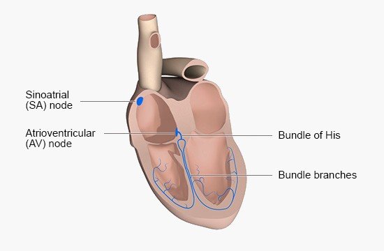
There is an area of cells known as the sinoatrial (SA) node in the right upper chamber of the heart. Also known as the sinus node, it is the main pacemaker for the heart rate. The SA node cells send out electrical signals at regular intervals, usually about 60 to 80 times per minute. The heart beats faster during physical exertion, fever, and mental stress such as anxiety – even up to 200 beats per minute (bpm) during very strenuous activities.
The atrioventricular (AV) node is located near the center of the heart, where the upper chambers (atria) and lower chambers (ventricles) meet. The AV node receives the signals sent from the SA node and then quickly passes them on to the lower chambers. It does that by sending them to the bundle of His. The cells in the AV node can also trigger signals themselves. But they only do that in certain situations, like if the SA node fails to work properly.
The bundle of His is a short strand of special muscle cells found in the wall that separates the left and right sides of the heart. It splits into two branches there. Known as the left and right bundle branches or “Tawara” branches, they lead to the lower end of the heart. They then split up into lots of thinner branches that reach throughout the heart muscle and pass on signals to both lower chambers. These fibers are called Purkinje fibers.
What do the electrical signals do?
When an electrical signal is sent out by the SA node, it first spreads out evenly down the muscles in the two upper heart chambers. No special electrical pathways are needed for that. This causes the muscles of the upper chambers to contract (squeeze), pumping the blood into the lower chambers.
While the upper chambers are squeezing, the electrical signal reaches the AV node. It then immediately spreads down the lower chambers – along the bundle of His, the bundle branches and the Purkinje fibers. This makes the muscle cells in the lower heart chambers squeeze strongly at the same time. They pump the fresh blood into the body and the used blood (returning from the rest of the body) into the lungs.
After one heartbeat, all of the heart muscle cells relax again and wait for the next signal.
Brandes R, Lang F, Schmidt R. Physiologie des Menschen: mit Pathophysiologie. Berlin: Springer; 2019.
Pschyrembel Online. 2025.
IQWiG health information is written with the aim of helping people understand the advantages and disadvantages of the main treatment options and health care services.
Because IQWiG is a German institute, some of the information provided here is specific to the German health care system. The suitability of any of the described options in an individual case can be determined by talking to a doctor. informedhealth.org can provide support for talks with doctors and other medical professionals, but cannot replace them. We do not offer individual consultations.
Our information is based on the results of good-quality studies. It is written by a team of health care professionals, scientists and editors, and reviewed by external experts. You can find a detailed description of how our health information is produced and updated in our methods.
Stay informed
Subscribe to our newsletter or newsfeed. You can find our growing collection of films on YouTube.

