Surgery for treating retinal detachment

Photo of a doctor and patient

If the has detached, fast surgery is needed to save or improve vision again. There are different procedures for reattaching the . Sometimes several procedures are needed.

Retinal detachment is where the is no longer in contact with the inside of the eyeball. That’s usually caused by small tears in the . The gel-like fluid from the vitreous body can pass through them and collect beneath the , meaning the is no longer supplied with nutrients from the layer below. That can permanently damage the in the .

Retinal detachment is a medical emergency, and surgery is usually the only treatment that makes sense. In most people, the can then be reattached. That usually means that the affected eye can be prevented from going blind and vision can be improved again.

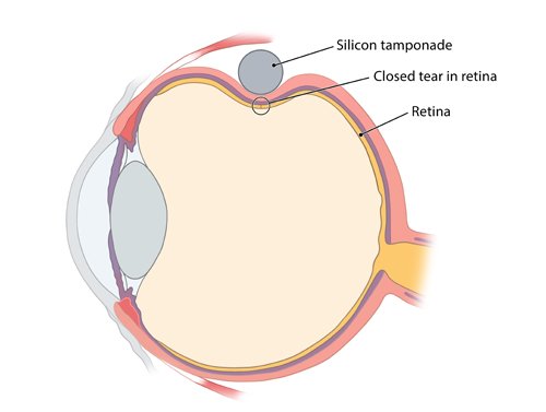
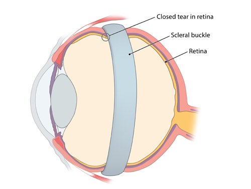
The two most common surgical procedures for reattaching the scleral buckling and removal of the vitreous body (vitrectomy). Which procedure makes most sense depends on a number of factors, including the size and the duration of the detachment. Sometimes, both methods are even combined.

What does scleral buckling involve?

In one of these procedures, the surgeon sews a silicone tamponade onto the outside of the eyeball where the retinal tear is. That then indents the wall of the eyeball, pressing the back onto the layer below. They are then “glued” back together using a laser or cryoprobe.

Alternatively, a silicone strip (scleral buckle) is wrapped around the eyeball in a ring pattern. That indents it all the way around, and several holes in the can be closed with one procedure.

Illustration: Scleral buckling procedures: Scleral buckle

Over time, the fluid under the is reabsorbed by the surrounding tissue. Sometimes, it can be sucked away during the operation using a very thin probe.

Vision is still impaired for a while after the procedure. It takes about 30 to 90 minutes to perform the operation. It’s performed in hospital under general or local anesthetic.

What does vitrectomy involve?

When removing the vitreous body (vitrectomy), the surgeon inserts fine instruments into the eye, sucks the vitreous body up, and replaces it first with silicone oil or a special gas. That presses the and the layer below it back together. The surgeon then “glues” the layers to one another using laser or cryotherapy.

It might be important to keep your head in a certain position for a few days after surgery, depending on where the was detached. Your doctor will tell you what you need to know after the operation.

The gas is gradually transported away via the blood vessels and replaced by the body’s own fluid. Depending on which gas was used, that can take 2 weeks to 3 months. Vision is very restricted in the affected eye during that time.

If silicone oil is used, it is usually sucked up during another procedure. The eyeball fills up with the body’s own fluid again afterwards.

Which gas or oil is used depends on a number of factors such as how big the holes are or how quickly you need to be active again or if you have to travel: If the vitreous body is replaced with silicone oil, vision is temporarily still somewhat impaired. If gas is used, vision is severely impaired at first. You also can’t take any flights or spend time in the mountains for the first few weeks after the procedure if gas is used. That’s because the gas expands the higher up you are and the lower the air pressure is, which can damage the eye.

You also have to stay in the hospital for this procedure. It is performed using general or local anesthetic and takes about 30 to 90 minutes.

Which procedure is suitable for you?

Your doctor will advise you about which procedure is best in your personal situation. A number of aspects are taken into account, such as

  • The size, number and location of the holes in the : Scleral buckling procedures are better suited for one or just a few holes. On the other hand, vitreous body removal (vitrectomy) is more commonly used for if the holes are big or there are many of them. Vitrectomy can also be a better option if the middle of the is damaged near to or on the eye’s focal point ().
  • Medical history: If your eye has already been operated on and an artificial lens was fitted or there was bleeding, vitrectomy is probably a better option.
  • The cause of the retinal detachment: Not all detached retinas are caused by tears. If it is caused by a build-up of fluid, vitrectomy is usually needed. If the has detached due to another disease such as a tumor, it’s important to also treat that.

How effective is surgery?

The can be reattached during surgery in most people and they can see more clearly and with better contrast again. This is true for both procedures. But vision is often no longer as good as before the detached . How well you will be able to see after surgery depends on various factors such as the position, size and duration of the detached .

Good to know:

Studies suggest that the can be reattached in about 95 out of 100 people. It can sometimes take several operations.

The detaches again in the same eye in the three years after the operation in about 20 to 30 out of 100 people. Another procedure is then necessary.

What are the possible complications?

Complications that can impair vision can occur during or after surgery in either procedure. Treatment with medications or another procedure might be needed if that happens. But permanent effects can usually be avoided.

The most common complications are:

After scleral buckling it’s possible that you might see double for a while or have difficulties moving your eye, like when tracking an object. But these problems usually go away by themselves after a few weeks or months.

New holes can form in the during vitrectomy. They can usually be closed up again during the surgery.

In rare cases, serious complications such as infections, inflammations or bleeding can occur during both procedures.

Important:

Quickly seek medical attention at an eye doctor’s or in an emergency room if you have severe pain or any worsening in eyesight after the procedure. Symptoms like that may be a sign of complications.

How do the advantages and disadvantages of the procedures compare?

Studies suggest that scleral buckling and vitrectomy are equally well suited for reattaching the and improving eyesight again. But they also find pointers that there are differences between some aspects of the procedures. For instance,

  • the studies found that it was less common for the to become detached again following vitrectomy. And fewer operations were needed to treat the retinal damage.
  • Different complications occurred: New tears in the only occurred during vitrectomy. Cataracts also developed more frequently after those operations.

But most of the studies only lasted a few months and looked at a small number of participants. That is why it isn’t yet possible to say whether one or other of the procedures is better.

Things to be aware of after surgery

You will usually stay in the hospital for a few days after surgery. Regular check-ups with an eye doctor are then needed to check things like eye pressure. It might be important to keep your head in a certain position for the first few days after the operation. Your doctor can give you more detailed information about that.

Doctors recommend eye drops or eye ointments to support the healing process. They can also prescribe painkillers if needed.

Your eye might be painful, swollen or red directly after surgery. Because it’s still sensitive, it’s important

  • not to rub your eyes
  • to make sure that no water, shampoo or dirt gets into the eye, and
  • not to do any physical exertion such as jogging or lifting heavy weights, or go to the sauna or swimming pool for about two weeks.
  • not to read or do any screen work for up to about two weeks to avoid the jolting movements when the eye moves from line to line. But it’s okay to watch television.

Your eyesight will be significantly worse for up to two weeks, especially if the eye was filled with gas after vitrectomy. That can affect your everyday life. You will only be able to drive again once your eye specialist has checked that your eyesight is good enough.

Deutsche Ophthalmologische Gesellschaft (DOG), Retinologische Gesellschaft (RG), Berufsverband der Augenärzte Deutschlands (BVA). Risikofaktoren und Prophylaxe der rhegmatogenen Netzhautablösung bei Erwachsenen (S1-Leitlinie). AWMF-Registernr.: 045-025. 2022.

Feltgen N, Walter P. Rhegmatogenous retinal detachment – an ophthalmologic emergency. Dtsch Arztebl Int 2014; 111(1-2): 12-21; quiz 22.

Gelston CD, Deitz GA. Eye Emergencies. Am Fam Physician 2020; 102(9): 539-545.

Lang GK. Augenheilkunde. Stuttgart: Thieme; 2014.

Pschyrembel Online. 2023.

Schwartz SG, Flynn HW, Wang X et al. Tamponade in surgery for retinal detachment associated with proliferative vitreoretinopathy. Cochrane Database Syst Rev 2020; (5): CD006126.

Sena DF, Kilian R, Liu SH et al. Pneumatic retinopexy versus scleral buckle for repairing simple rhegmatogenous retinal detachments. Cochrane Database Syst Rev 2021; (11): CD008350.

Znaor L, Medic A, Binder S et al. Pars plana vitrectomy versus scleral buckling for repairing simple rhegmatogenous retinal detachments. Cochrane Database Syst Rev 2019; (3): CD009562.

IQWiG health information is written with the aim of helping people understand the advantages and disadvantages of the main treatment options and health care services.

Because IQWiG is a German institute, some of the information provided here is specific to the German health care system. The suitability of any of the described options in an individual case can be determined by talking to a doctor. informedhealth.org can provide support for talks with doctors and other medical professionals, but cannot replace them. We do not offer individual consultations.

Our information is based on the results of good-quality studies. It is written by a team of health care professionals, scientists and editors, and reviewed by external experts. You can find a detailed description of how our health information is produced and updated in our methods.

Comment on this page

What would you like to share with us?

We welcome any feedback and ideas - either via our form or by gi-kontakt@iqwig.de. We will review, but not publish, your ratings and comments. Your information will of course be treated confidentially. Fields marked with an asterisk (*) are required fields.

Please note that we do not provide individual advice on matters of health. You can read about where to find help and support in Germany in our information “How can I find self-help groups and information centers?

Über diese Seite

Created on November 12, 2024

Next planned update: 2027

Publisher:

Institute for Quality and Efficiency in Health Care (IQWiG, Germany)

Stay informed

Subscribe to our newsletter or newsfeed. You can find our growing collection of films on YouTube.