Retinal detachment

At a glance

  • Retinal detachment (detached retina) is a rare and serious eye condition.
  • The retina comes loose from the inside of the eyeball and is then damaged.
  • Typical signs of retinal detachment include flashes of light and shady areas or dark patches in your vision.
  • It is important to see an eye specialist as soon as possible if you notice symptoms like that.
  • Retinal detachment is a medical emergency.
  • If it is detected early on and surgery is performed, vision loss can often be prevented.

Introduction

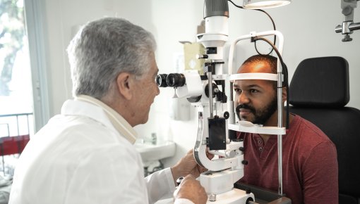
Photo of a patient and a doctor during an eye examination

The is the inner lining of the eyeball. It contains millions of that enable us to see colors and light. But if little tears or holes form in the , it can detach from the layer below.

Retinal detachment can cause a number of vision problems, such as flashes of light, streaks, blacks dots, shady patches in your field of vision, or loss of sight in one eye.

It is important to see an eye specialist (ophthalmologist) as soon as possible if you have symptoms like that. Retinal detachment is a medical emergency and needs to be treated as quickly as possible. Otherwise, the may continue to detach and the affected eye could go blind. Surgery can be used to re-attach the and improve vision.

Symptoms

Retinal detachment doesn't cause any pain. Even in its early stages it is noticeable because of the visual problems it causes, including:

  • Flashes of light that are sometimes visible even when you close your eyes
  • Bright streaks, rings or black dots which move when you look around (“floaters”)
  • Dark patches that flow across your field of vision
  • Little falling black dots (“black rain”)
  • Shadows that cover the field of vision like curtains being drawn from the outside to the inside lasting several hours or days

Things like exactly where the is detaching will determine which of these symptoms develop. If the middle of the () is affected, your vision can become blurry or distorted.

Illustration: Possible signs of retinal detachment: Flashes of light
Illustration: Bright streaks, rings or black dots which move when you look around (“floaters”)
Illustration: Dark patches that flow across your field of vision
Illustration: Shadows that cover the field of vision
Illustration: Blurry or distorted vision

Causes

The most common cause of retinal detachment is a torn .

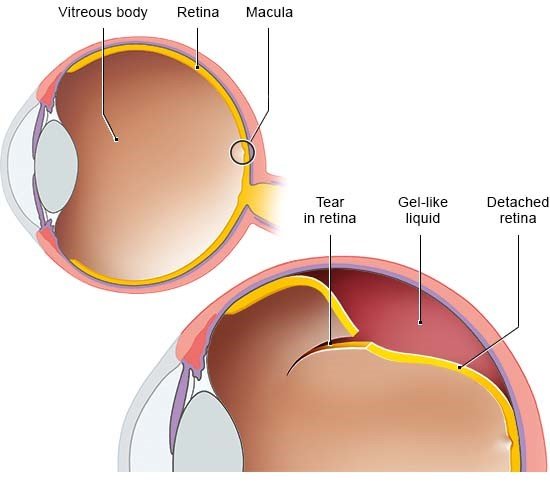
The is loosely attached to a layer below it, which supplies the with nutrients. The gel-like liquid in the middle of the eye (vitreous body) can get under the through a tear or a hole in the . That pushes the away from the layer beneath, meaning that the in the no longer get enough nutrients. That leads to the typical symptoms of retinal detachment and causes permanent damage to the .

Image: Left: healthy eye, right: eye with retinal detachment caused by a tear

Not all detached retinas are caused by tears. They can sometimes be caused by scars on the or a build-up of fluids resulting from other medical conditions, but both cases are much less common.

Risk factors

There is an increased risk of detached caused by tears in people who have:

  • moderately to severe nearsightedness,
  • close relatives with detached (genetic predisposition),
  • already had eye surgery, e.g. for cataracts or far or nearsightedness,
  • have injured their eye in the past, perhaps when doing martial arts or ball sports.

In the years following retinal detachment in one eye, the of the other eye also detaches in up to 20 out of 100 people.

Some people have retinal damage as a result of diabetes (diabetic retinopathy). Retinal detachment is more common in those people.

Prevalence

Experts estimate that retinal detachment affects about 10 to 25 out of 100,000 people in central Europe per year.

It becomes increasingly common in older age: About 3% of all people experience retinal detachment by the age of 85. It is more common in men than in women. But children and people under the age of 30 years are less commonly affected.

Outlook and effects

If the has detached, a swift and surgery are key to saving or restoring vision. It is possible that the affected eye will go blind without surgery.

The can usually be re-attached. Sometimes, several procedures are needed. How good your eyesight will be afterwards depends on things such as where exactly the detached from and how severe the damage already was.

Diagnosis

Your eye specialist will first ask you about your symptoms and any other conditions or injuries you might have. Your eye will then be closely examined. A vision test is sometimes done first to check your eyesight. The doctor will look into your eyes using a special microscope called a slit lamp.

This is followed by an examination of the back of the eye to take a closer look at the there. You will be given eye drops beforehand to dilate (widen) the pupils. The doctor will shine a special device called an ophthalmoscope directly into your eye to make the visible. They can then see if there are any tears or detachments. Your eyes will be more sensitive to light and your vision will be a little blurry for a while after the examination.

Other examinations such as ultrasound scans might also be done afterwards.

Prevention

Age-related retinal detachment often can’t be avoided with preventive measures. But it’s important to see an eye specialist quickly if you notice changes in your eyesight. Permanent loss of vision can often be prevented if detached is detected and treated early on.

Regular check-ups are important if you have an increased risk of retinal detachment, for example due to nearsightedness. Pre-stages of retinal detachment such as tears can be treated at your doctor’s office to prevent full detachment.

There are two options: laser treatment (thermal laser coagulation) or cold therapy (cryocoagulation). Heat or cold is then used to attach the back onto the lower layer around the tear or hole. The tears are then still there, but can’t get any bigger.

The risk of eye injuries and related retinal detachment can be reduced by wearing safety goggles when doing certain activities, like work that involves sawing, drilling or hammering, but also contact and ball sports such as hockey and squash.

Treatment

Retinal detachment usually calls for fast surgery in hospital. The two most common types of operation in Germany are:

  • Scleral buckling: A silicone seal or a silicone ring is used to indent the eyeball from the outside so that the is in contact with the layer below again.
  • Surgery to remove the vitreous body (vitrectomy): The gel-like vitreous body is replaced by gas or silicone oil. The two layers are then pressed together again. The gas is then replaced by the body’s natural fluid over time. But another procedure is needed to remove the silicone oil.

The choice of procedure depends on things such as where and how severely the is damaged, but also whether the eye has already been operated on before. It is best to talk to your doctor about which procedure makes the most sense in your individual situation.

They can also give you valuable tips about what you have to be aware of before and after the operation. For example, it’s important to avoid quick head and eye movements to prevent the from detaching more quickly.

It is usually possible to surgically reattach the detached . That often also improves eyesight in that area. But if the middle of the () is affected, vision usually remains permanently impaired, with blurred vision and less contrast.

Everyday life

Being told you have retinal tears by an eye specialist can be worrying. Even if you don’t have any vision problems yet, knowing that you have an increased risk of retinal detachment can be unsettling and distressing.

People with sudden, severe vision problems due to retinal detachment are often scared of the possible serious consequences. Above all, they are often very worried about going blind. But that can usually be prevented if it is treated early enough.

No longer being able to see very well due to retinal detachment can affect your everyday life. Certain strategies and adjustments can then help you to cope better, such as:

  • Rearranging things at home: Contrasts, color accentuations and good lighting can make it easy to find your way around at home. For instance, you can put brighter colors on corners or light switches.
  • Using aids at work and in your free time: There are lots of useful aids, such as magnifying glasses and voice-controlled household devices. Smartphone and tablet apps can also come in handy, e.g. for reading out text or identifying objects. You can get information at your doctor’s office or from your health insurer about useful aids and which costs might be covered. Company doctors are responsible for such topics at the workplace.
  • Talking to other people who have had retinal detachment: Many people with vision problems find it helpful to talk to other people with the same condition, like at a support group. Real-life stories can give you an idea of how other people cope with the disease and its effects.
  • Support from family and friends: People with vision problems are more reliant on help from others. They can help you to stay as active as possible. It can be a relief to put your trust in them and talk to them about your fears and concerns.

Further information

Eye doctors are usually the first point of contact for eye conditions. In our topic "Health care in Germany" you can read about how to find the right doctor – and our list of questions can help you to prepare for your visit to the doctor.

In Germany there are various places people with eye conditions can turn to for help, including support groups and information centers. Our list might help you to find the support you’re looking for.

American Optometric Association. Care of the Patient with Retinal Detachment And Related Peripheral Vitreoretinal Disease. 2004.

Bechrakis NE, Dimmer A. Rhegmatogene Netzhautablösung Epidemiologie und Risikofaktoren. Ophthalmologe 2018; 115: 163-178.

D'Amico DJ. Clinical practice. Primary retinal detachment. N Engl J Med 2008; 359(22): 2346-2354.

Deutsche Ophthalmologische Gesellschaft (DOG), Retinologische Gesellschaft (RG), Berufsverband der Augenärzte Deutschlands (BVA). Risikofaktoren und Prophylaxe der rhegmatogenen Netzhautablösung bei Erwachsenen (S1-Leitlinie). AWMF-Registernr.: 045-025. 2022.

Feltgen N, Walter P. Rhegmatogenous retinal detachment – an ophthalmologic emergency. Dtsch Arztebl Int 2014; 111(1-2): 12-21; quiz 22.

Gelston CD, Deitz GA. Eye Emergencies. Am Fam Physician 2020; 102(9): 539-545.

Kang HK, Luff AJ. Management of retinal detachment: a guide for non-ophthalmologists. BMJ 2008; 336(7655): 1235-1240.

Lang GK. Augenheilkunde. Stuttgart: Thieme; 2014.

Sena DF, Kilian R, Liu SH et al. Pneumatic retinopexy versus scleral buckle for repairing simple rhegmatogenous retinal detachments. Cochrane Database Syst Rev 2021; (11): CD008350.

Ullrich M, Zwickl H, Findl O. Incidence of rhegmatogenous retinal detachment in myopic phakic eyes. J Cataract Refract Surg 2021; 47(4): 533-541.

IQWiG health information is written with the aim of helping people understand the advantages and disadvantages of the main treatment options and health care services.

Because IQWiG is a German institute, some of the information provided here is specific to the German health care system. The suitability of any of the described options in an individual case can be determined by talking to a doctor. informedhealth.org can provide support for talks with doctors and other medical professionals, but cannot replace them. We do not offer individual consultations.

Our information is based on the results of good-quality studies. It is written by a team of health care professionals, scientists and editors, and reviewed by external experts. You can find a detailed description of how our health information is produced and updated in our methods.
Comment on this page

What would you like to share with us?

We welcome any feedback and ideas - either via our form or by gi-kontakt@iqwig.de. We will review, but not publish, your ratings and comments. Your information will of course be treated confidentially. Fields marked with an asterisk (*) are required fields.

Please note that we do not provide individual advice on matters of health. You can read about where to find help and support in Germany in our information “How can I find self-help groups and information centers?

Über diese Seite

Created on November 12, 2024

Next planned update: 2027

Publisher:

Institute for Quality and Efficiency in Health Care (IQWiG, Germany)

Stay informed

Subscribe to our newsletter or newsfeed. You can find our growing collection of films on YouTube.