How do our feet work?

When you walk, your feet carry the entire weight of your body and keep you balanced at the same time. So the strain on them is among the highest loads your body has to carry.

Most of your body weight rests on the heels and balls of your feet. Your leg and foot muscles make small stabilizing movements to ensure you don’t lose your balance when standing or walking.

Bones and joints

The human foot consists of 26 bones in total:

  • 7 tarsals (at the back of the foot)
  • 5 metatarsals (in the middle of the foot)
  • 14 phalanges (in the toes)

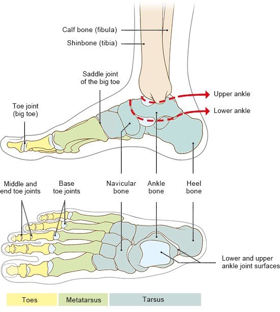
They are all connected to one another by joints and ligaments. The foot can be divided into three sections based on the different joints. These sections are called the tarsus (including the ankle), the metatarsus and the toes.

The ankle connects the bones in the lower leg to the bones of the foot. It can be divided into two parts: the upper and lower ankle.

Illustration: Bones and joints in the foot

Tarsus and ankle

The seven tarsal bones are held together tightly by ligaments, and are more or less fixed in place. They are grouped in two rows – one with three bones and one with four. One of these bones – the ankle bone (talus) – forms part of the upper ankle joint along with the shinbone (tibia) and calf bone (fibula). The shin and calf bones are separated from the ankle bone by a layer of cartilage. The ankle bone is also connected to the tarsal bones and the heel bone (calcaneous). The lower ankle joint is found between the heel bone, ankle bone and navicular bone.

The upper ankle allows you to move your feet upwards, downwards, and a little to the side. The lower ankle connects the ankle bone to the bones in the tarsus and the heel bone. It doesn't move as much as the upper ankle. The lower ankle enables you to tilt your feet sideways slightly and turn them inward and outward.

Metatarsus

The second row of tarsals is connected to the metatarsus. This consists of five long bones. You can feel them quite clearly along the top of your foot. One of these metatarsal bones and the bone at the base of the big toe come together to form a saddle joint, which gives big toes their particular flexibility.

Toes

The freely movable part of your foot is made up of five toes. Each toe has three bones – apart from the big toe, which only has two. They also have three joints: at the base, in the middle and towards the end of the toe. The base joints are ball-and-socket joints, which allow you to move your toes up and down, and to spread them out a little too. The middle joints are hinge joints and can only move in one direction, meaning that they can only stretch or bend the toes.

The arch of the foot

For your feet to stay supple and protect the sensitive muscles, tendons, blood vessels and nerves inside them, they have to be able to absorb impact. Normally, the soles of the feet do this job by forming a kind of arch between the heel and ball of the foot instead of resting flat on the ground.

The feet each have a longitudinal arch (along the length of the foot) and a transverse arch (from one side to the other). Muscles stretched along and across the foot pull the bones together to create an arch that is constantly under tension. When the foot has to carry a load – for instance, when you take a step – the muscles loosen slightly, allowing the arch to absorb the weight of your body. In a healthy foot, only the heel, ball of the foot and toes are in contact with the ground. A layer of fat beneath them provides protection. The metatarsus is given extra protection by a thick band of connective tissue called the “plantar fascia.” This tissue plays an important role in walking and standing, and tightens when the foot is placed on the ground.

Muscles

The foot’s muscle structure is extremely complex and consists of more than 30 muscles. Most of your foot movements are also controlled by muscles in your lower leg. It is only the thin tendons of these muscles (known as extensor and flexor muscles) that run down into the actual foot. The tendons of the extensor muscles run along the top of the foot to the ends of the toes, whilst the flexor tendons start at the heel or run along the sole of the foot to the toes.

Short foot muscles

The short foot muscles are found between the metatarsal bones. They help you spread out, bend (flex) and stretch (extend) your toes, but their main task is to tighten the arch of the foot.

Unlike our fingers, our toes aren't very flexible. Apart from the big and little ones, it’s almost impossible to move them independently of one another. There are a number of flexor muscles along the sole of your feet that move the three middle toes at the same time, and another muscle along the top of your feet that stretches them.

Several muscles in the soles of your feet are mainly there to support the arches. They pull the foot into a bow shape by pulling the forefoot and tarsus together. Most of these muscles run along the length of the foot because they also bend the toes. They create the arch between the front and back of the foot. The arch that goes from one side of the foot to the other is mainly supported by the muscle that’s responsible for pulling the big toe inward – the only muscle that doesn't run along the length of the foot.

When you raise your arch, the muscles in your foot are supported by the muscles and tendons in your lower leg.

Blood supply

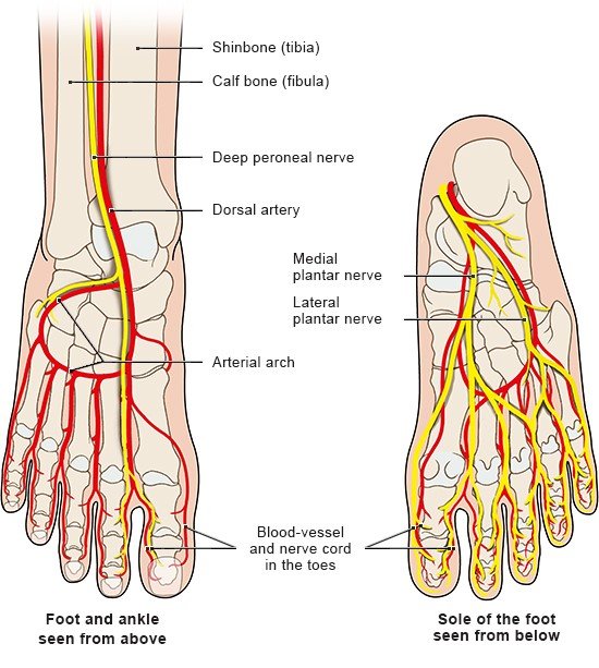
Two main arteries supply blood to the foot: one at the top of the foot (dorsal artery) and one in the sole (plantar artery) of the foot. The dorsal artery runs along the inner side of the top of the foot, along the big toe’s extensor muscle. It ends in an arch, from which other arteries branch off to supply blood to the upper side of your toes.

The plantar artery divides into two branches in the heel area. One runs along the inner side of your sole (medial plantar artery), the other in an arch shape on the outer side (lateral plantar artery). There are also arteries leading off from the lateral plantar artery into your toes. The arterial arches along the top and the sole of your foot are connected to one another.

Illustration: Blood vessels and nerves in the foot

Nerves

The muscles and skin of the foot are supplied by three nerves:

  • the deep peroneal nerve,
  • the medial plantar nerve, and
  • the lateral plantar nerve.

The deep peroneal nerve runs along the dorsal artery and moves the muscles that stretch your toes. One branch runs to a point between the big toe and the second toe, where it then splits into two other branches. The deep peroneal nerve also picks up sensory information from the skin along the top of the foot.

The medial (inner) and lateral (outer) plantar nerves run along the sole of your foot. The medial plantar nerve already splits near the heel, forming several branches along the sole of the foot. It moves the flexor muscles of your first three toes and, to a certain extent, the fourth one. It also picks up skin sensations from the sole of your foot up to the fourth toe.

The lateral plantar nerve follows the lateral plantar artery up to the little toe. It moves some of the fourth toe’s flexor muscles and the little toe’s flexor muscle. It also picks up sensory information from the skin in this area and passes it on to your brain.

Lippert H. Lehrbuch Anatomie. München: Urban und Fischer; 2017.

Menche N (Ed). Biologie Anatomie Physiologie. München: Urban und Fischer; 2016.

IQWiG health information is written with the aim of helping people understand the advantages and disadvantages of the main treatment options and health care services.

Because IQWiG is a German institute, some of the information provided here is specific to the German health care system. The suitability of any of the described options in an individual case can be determined by talking to a doctor. informedhealth.org can provide support for talks with doctors and other medical professionals, but cannot replace them. We do not offer individual consultations.

Our information is based on the results of good-quality studies. It is written by a team of health care professionals, scientists and editors, and reviewed by external experts. You can find a detailed description of how our health information is produced and updated in our methods.

Comment on this page

What would you like to share with us?

We welcome any feedback and ideas - either via our form or by gi-kontakt@iqwig.de. We will review, but not publish, your ratings and comments. Your information will of course be treated confidentially. Fields marked with an asterisk (*) are required fields.

Please note that we do not provide individual advice on matters of health. You can read about where to find help and support in Germany in our information “How can I find self-help groups and information centers?

Über diese Seite

Created on October 20, 2021

Next planned update: 2024

Publisher:

Institute for Quality and Efficiency in Health Care (IQWiG, Germany)

How we keep you informed

Follow us on Twitter or subscribe to our newsletter or newsfeed. You can find all of our films online on YouTube.