How does skin work?

Even at its thickest point, our skin is only a few millimeters thick. But it is still our heaviest and largest organ, making up about one seventh of our body weight: Depending on your height and body mass, it weighs between 3.5 and 10 kilograms (7.5 and 22 pounds) and has a surface area of 1.5 to 2 square meters.

The thickness of your skin varies depending on the part of the body it covers and how much it is used, but also depends on your age and sex: Older people generally have thinner skin than younger people do, and men generally have thicker skin than women do.

What does skin do?

The skin is our central sensory organ, and it carries out a number of tasks. It is a stable but flexible outer covering that acts as barrier, protecting your body from harmful things in the outside world such as moisture, the cold and sun rays, as well as germs and toxic substances.

Skin also plays an important role in regulating your body temperature. It helps prevent dehydration and protects you from the negative effects of too much heat or cold. And it allows your body to feel sensations such as warmth, cold, pressure, itching and pain.

Skin also functions as a large storeroom for the body: The deepest layer of skin can store water, fat and metabolic products. And it produces that are important for the whole body.

Just looking at someone’s skin can already tell you a lot – for instance, about their age and health. Changes in skin color or structure can be a sign of a medical condition. Regardless of their general skin type, people with too few red blood cells in their blood may look pale, and people who have hepatitis have yellowish skin. These changes may be less apparent in darker skin.

If skin is injured, the blood supply to the skin increases in order to deliver various substances to the wound so it is better protected from infections and can heal faster. Later on, new cells are produced to form new skin and blood vessels. Depending on how deep the wound is, it heals with or without a scar.

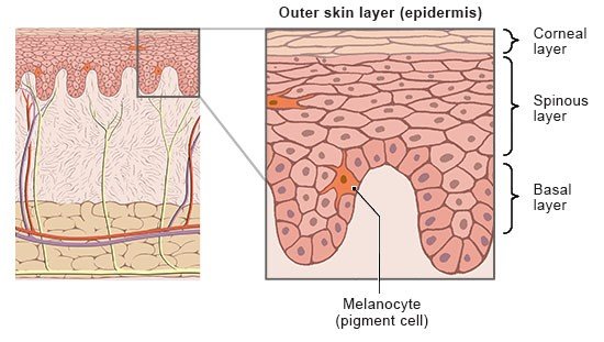
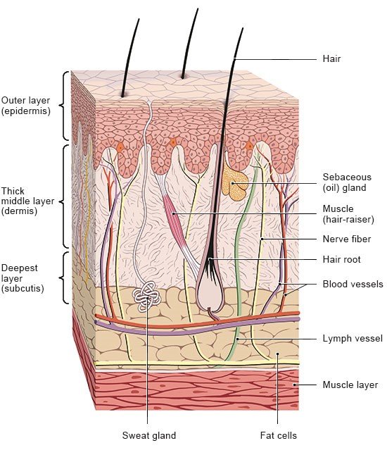
To be able to do all of these things, skin consists of three different layers: the outer layer (epidermis), the middle layer (dermis) and the deepest layer (subcutis).

What is the epidermis?

The outermost layer of skin which you can see is called the epidermis. It varies in thickness on different parts of the body: For instance, it is only 0.3 millimeters thick on your elbows and the back of your knees, and up to 4 millimeters thick on other parts of your body such as the soles of your feet and palms of your hands.

The epidermis is mostly made up of cells that produce keratin (keratinocytes), and it is constantly renewing itself: New cells are made in the lower layers of the epidermis. These move to the surface within four weeks, where they harden and are then die off. The hardened keratinocytes are packed closely together and seal the skin off from the outside environment. They are shed as tiny flakes or are rubbed off. The constant renewal process of the cells makes up for what is lost on the surface.

The cells in the epidermis grow faster in response to pressure or rubbing. The amount of skin flakes that are shed remains the same, though. As a result, the layer of hardened skin on the surface gradually becomes thicker and a callus develops. The skin does this to protect itself – to better withstand pressure and rubbing.

The anatomical illustration shows the position of the epidermis and provides a detailed view of its structure.

Only rarely is the balance of new cell production and old cell shedding affected by illness. Examples include infections, autoimmune disorders or genetic diseases that cause increased growth of rough, scaly skin on the entire body.

The epidermis also contains other types of cells with special functions:

  • Melanocytes produce and store a black pigment called melanin. They produce more melanin when your skin is exposed to sunlight, which is why it becomes darker. This protects the skin from the sun’s harmful UV rays.
  • Lymphocytes and Langerhans cells play an important role in fighting germs. They “grab” the germs and take them to the nearest lymph node.
  • Merkel cells are special nerve cells in the skin that enable you to sense pressure.
The anatomical illustration shows the structure of the skin.

What does the dermis do?

Under the epidermis, firmly stuck to it, lies the middle layer of skin (the dermis). It is made up of a dense network of tough, elastic fibers. These make the skin strong and robust, while at the same time stretchy. If the skin is stretched a lot, for instance the skin covering the belly during pregnancy, tears in the dermis may result, which can be seen as light lines.

In places, the dermis bulges into the connective tissue that surrounds our muscles and bones and connects them with the skin.

The dermis contains a network of nerve fibers and very small blood vessels called capillaries. Nutrients and oxygen in the blood pass from the capillaries into cells. Another main function of the dermis is to help your body cool down if it gets too hot. It is also the skin layer that contains the most sensory (feeling) cells and sweat glands.

What does the subcutis do?

The subcutis (also known as the subcutaneous layer or hypodermis) is mostly made up of fat and connective tissue. In the subcutis, between the folds of dermis that bulge into it, there are tiny cavities. These cavities are filled with storage tissue made out of fat and water. The fat acts as a shock absorber, protecting bones and joints from blows or bumps. It serves as insulation too. What’s more, many hormones are produced in the fat cells of the subcutis. One example is vitamin D, which is an essential vitamin and is made when the skin is exposed to sunlight.

The subcutis and dermis both contain blood vessels and lymph vessels too, as well as other things like nerves, sweat glands, sebaceous (oil) glands, scent glands and hair roots.

Brandes R, Lang F, Schmidt R. Physiologie des Menschen: mit Pathophysiologie. Berlin: Springer; 2019.

Mayo Clinic. Anemia. 2025.

Mayo Clinic. Liver disease. 2025.

Menche N. Biologie Anatomie Physiologie. Munich: Urban und Fischer; 2023.

Moll I. Duale Reihe Dermatologie. Stuttgart: Thieme; 2016.

Pschyrembel Online. Haut. 2025.

Williams H, Bigby M, Herxheimer A et al. Evidence-Based Dermatology. Hoboken: Wiley; 2014.

IQWiG health information is written with the aim of helping people understand the advantages and disadvantages of the main treatment options and health care services.

Because IQWiG is a German institute, some of the information provided here is specific to the German health care system. The suitability of any of the described options in an individual case can be determined by talking to a doctor. informedhealth.org can provide support for talks with doctors and other medical professionals, but cannot replace them. We do not offer individual consultations.

Our information is based on the results of good-quality studies. It is written by a team of health care professionals, scientists and editors, and reviewed by external experts. You can find a detailed description of how our health information is produced and updated in our methods.

Comment on this page

What would you like to share with us?

We welcome any feedback and ideas - either via our form or by gi-kontakt@iqwig.de. We will review, but not publish, your ratings and comments. Your information will of course be treated confidentially. Fields marked with an asterisk (*) are required fields.

Please note that we do not provide individual advice on matters of health. You can read about where to find help and support in Germany in our information “How can I find self-help groups and information centers?

Über diese Seite

Updated on April 22, 2025

Next planned update: 2028

Publisher:

Institute for Quality and Efficiency in Health Care (IQWiG, Germany)

Stay informed

Subscribe to our newsletter or newsfeed. You can find our growing collection of films on YouTube.