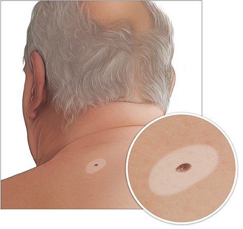
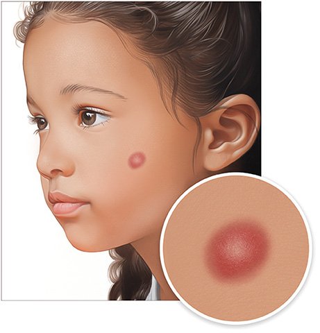
What do moles, birthmarks and other nevi look like?

Photo of a larger mole on the neck

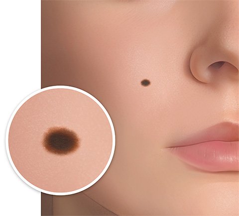
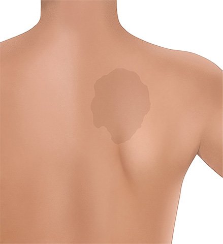
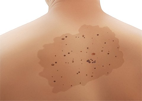
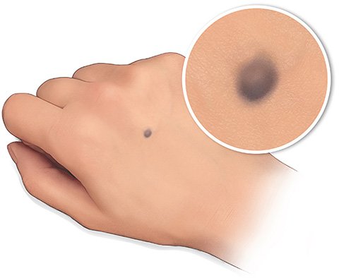
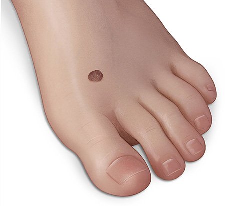
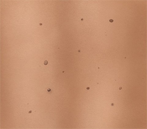
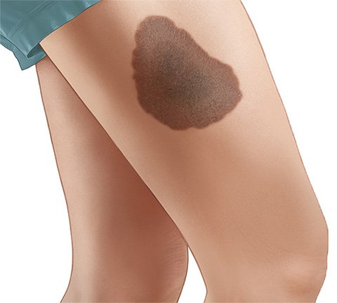
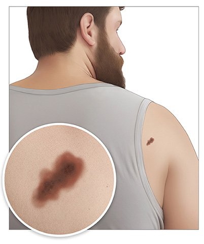
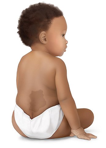
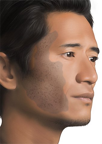
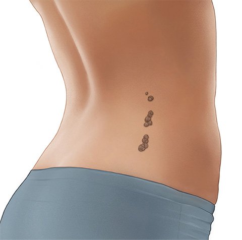
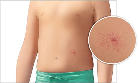
Moles and other nevi are typically just a few millimeters in size, oval or round in shape, and darker than the surrounding skin. But some are much bigger and have a different color or shape – such as reddish port-wine stains or slate gray nevi on the lower back.

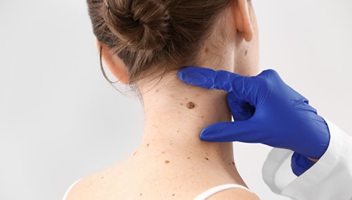
Many people have spots or areas of skin that are darker than the rest of their skin. Commonly known as moles, birthmarks or beauty spots, most of these are considered to be a nevus (plural: nevi) from a medical point of view.

Good to know:

Moles and other types of nevus are usually benign and only rarely turn into cancer. But it’s still a good idea to regularly check your skin and be aware of warning signs, like changes in color or shape.

What do pigmented nevi look like?

Pigmented nevi are made of cells that produce and store the dark-colored pigment melanin, and sometimes also release it into the surrounding cells. These melanin-producing cells are called melanocytes. If a lot of them build up in one place or some are especially active, the color of the skin there changes.

Pigmented nevi are more common on lighter skin than on darker skin. They are also more noticeable on lighter skin because of the brown color. There are different types of pigmented nevi:

  • Simple lentigines (Lentigo simplex) are brown to dark brown in color, and usually just a few millimeters in size. They are in the top layer of the skin or mucous membrane. Simple lentigines usually develop in childhood or the teenage years.
Illustration: A simple lentigo (Lentigo simplex)
  • Café au lait spots can be anywhere between the size of a fingernail and palm of the hand. They have a clear border. On lighter skin they're light brown in color – quite like the color of coffee with milk ("café au lait" in French). On dark skin they look a little darker than the surrounding skin. Café au lait spots are in the top layer of skin. They develop in children and teenagers. More than five of these spots on your body could be a sign of a genetic disorder such as neurofibromatosis (Von Recklinghausen's disease).
Illustration: A café au lait spot
  • Nevus spilus is quite rare. It is a mixture of a café au lait spot and scattered clusters of darker pigment cells.
Illustration: A nevus spilus
  • Blue nevi (Nevus coeruleus) are similar to simple lentigines, but they're more bluish, purple, or gray-brown in color because they're deeper in the skin. They can often be felt as a small “lump.” If at all, people usually only have one blue nevus (or very few) and it only appears in the course of their life. Blue nevi typically develop on the back of the hand or the forearm.
Illustration: A blue nevus (Nevus coeruleus)
  • Melanocytic nevi appear as small spots or larger patches, on their own or in groups. These common moles can be any shade of brown or even look almost black. They are often hairier than the surrounding skin. Some melanocytic nevi are already visible at birth. But they often only develop in childhood, and then disappear again in later life. They can change over that time and, for example, move from the upper layer of skin to lower layers. They then usually get lighter in color and become a little more raised, which can make them look like light warts.
Illustration: Single melanocytic nevus
Illustration: Group of several nevi
Illustration: Large melanocytic nevus


  • Dysplastic or atypical nevi are special types of melanocytic nevi that look different to other pigmented nevi: They are usually bigger than five millimeters and have an irregular edge. A dysplastic nevus can have a mixture of different colors in it, including pink, light brown, dark brown and black. The term “dysplastic” refers to the properties of the cells in the nevus. The risk of developing melanoma skin cancer is believed to be higher in people who have a lot of dysplastic nevi. This kind of cancer can develop from a dysplastic nevus, but it sometimes also develops on skin that used to be "normal."
Illustration: A dysplastic (atypical) nevus
  • Slate gray nevi (previously known as Mongolian spots) are already there at birth. They can be seen as a gray-blue patch on the lower back, tailbone area or buttocks. These patches mainly develop in children with darker skin and those of Asian origin. They usually go away again on their own after puberty.
Illustration: A slate gray nevus on a baby
  • A nevus of Ota is a relatively large, gray-blue patch on one side of the face, usually spreading over the temple and cheek. The eye is sometimes affected too, but that doesn’t impair your vision. These nevi are common in people of Asian descent. Some are already there from birth, and others only develop in puberty. They usually don't go away again.
Illustration: A nevus of Ota
  • Halo nevi are surrounded by a light, halo-like ring – probably because an autoimmune reaction in the body breaks down the pigment around the mole.
Illustration: A halo nevus with a light ring ("halo") around it
  • Spitz nevi tend to look like a reddish bump. They develop in children and teenagers. They are harmless, but can be easily confused with other, more serious skin changes.
Illustration: A Spitz nevus

What about nevi that aren't made of pigment cells?

Nevi can also develop from other skin cells, or from connective or fatty tissue. Even without the typical pigment cells, some are darker than the surrounding skin. If they develop from skin cells, they often form in groups and look as if they're arranged along a string.

Illustration: Nevi from skin cells

Dilated (wide) and unusually arranged blood vessels may also be visible as nevi – often just as red-purplish dots that appear with increasing age. But there are other types, too:

  • Nevus flammeus is known as a port-wine stain because of its bright reddish-pink color. It is sometimes a pale pink or purple-bluish color instead. This kind of birthmark is often less noticeable on dark skin than on light skin. Some are as small as a lentil, while others cover larger areas of skin, such as one side of the face. They are usually present at birth or appear in very early childhood. When they develop on the back of the neck, they're called stork bites. Those on the forehead or back of the neck may get paler over time. They usually stay the same in other places, though. Port-wine stains may be a sign of a genetic disease.

    It can be difficult to tell the difference between port-wine stains and hemangiomas (non-cancerous growths of blood vessels).
Illustration: Child with nevus flammeus on the back of their neck (stork bite)
  • A spider nevus looks like a red dot surrounded by thin red lines extending from it, like spider’s legs. Spider nevi can develop in children or suddenly during pregnancy. They are more common in people with liver disease.
Illustration: A spider nevus

Bendick C. Besonderheiten der nichtweißen Haut. In: Braun-Falco's Dermatologie, Venerologie und Allergologie. Berlin: Springer; 2017.

Holm N, Burgdorf WH. Hauterkrankungen bei dunkelpigmentierten Menschen. In: Plewig G, Degitz K (Ed). Fortschritte der praktischen Dermatologie und Venerologie, vol 17. Berlin: Springer; 2001.

Leitlinienprogramm Onkologie (Deutsche Krebsgesellschaft, Deutsche Krebshilfe, AWMF). Prävention von Hautkrebs (S3-Leitlinie). AWMF-Registernr.: 032-052OL. 2021.

Moll I. Duale Reihe Dermatologie. Stuttgart: Thieme; 2016.

Pschyrembel Online. 2023.

Tronnier M. Melanotische Flecke und melanozytäre Nävi. In: Braun-Falco's Dermatologie, Venerologie und Allergologie. Springer; 2018.

IQWiG health information is written with the aim of helping people understand the advantages and disadvantages of the main treatment options and health care services.

Because IQWiG is a German institute, some of the information provided here is specific to the German health care system. The suitability of any of the described options in an individual case can be determined by talking to a doctor. informedhealth.org can provide support for talks with doctors and other medical professionals, but cannot replace them. We do not offer individual consultations.

Our information is based on the results of good-quality studies. It is written by a team of health care professionals, scientists and editors, and reviewed by external experts. You can find a detailed description of how our health information is produced and updated in our methods.

Comment on this page

What would you like to share with us?

We welcome any feedback and ideas - either via our form or by gi-kontakt@iqwig.de. We will review, but not publish, your ratings and comments. Your information will of course be treated confidentially. Fields marked with an asterisk (*) are required fields.

Please note that we do not provide individual advice on matters of health. You can read about where to find help and support in Germany in our information “How can I find self-help groups and information centers?

Über diese Seite

Created on February 28, 2024

Next planned update: 2027

Publisher:

Institute for Quality and Efficiency in Health Care (IQWiG, Germany)

How we keep you informed

Follow us on Twitter or subscribe to our newsletter or newsfeed. You can find all of our films online on YouTube.