How does the heart work?

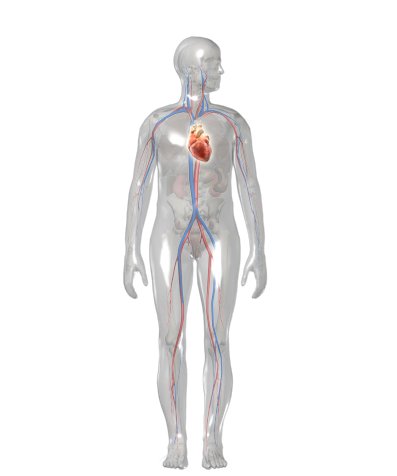
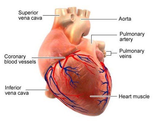
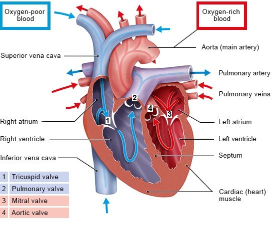
The heart provides the body’s organs and tissues with a constant supply of blood – and with it vital oxygen and nutrients. You can think of the heart as a central pump that keeps the blood circulating around the body.

In adults, the heart beats about 60 to 80 times per minute at rest. With every heartbeat, it pumps blood through the body. When you do strenuous physical activities, your heart beats at a faster rate and blood flows more quickly through your body. The blood can then absorb more oxygen from the lungs per minute in order to supply the body's cells with enough oxygen.

The illustration shows a human body from the front and the location of the heart within it. It is roughly in the middle of the chest.

Your heart is about the same size as your fist and weighs around 300 g (about 0.7 pounds). In people who do a lot of endurance sports, it can weigh up to 500 g (about 1.1 pounds). The heart is located more or less in the middle of the chest, slightly to the left, behind the breastbone. You can normally feel someone’s heart beat if you put your hand on their chest.

The illustration shows the heart and the coronary arteries, which run directly along the heart muscle and supply it with oxygen and nutrients.

How does blood flow through the heart?

The heart is a hollow muscle. A wall through the middle (known as the septum) divides it into two halves. Each half has two chambers called the atrium and ventricle. The left ventricle pumps oxygen-rich blood out of the heart and into the body (systemic circulation) through the main artery, called the aorta. The first blood vessels that branch off from the aorta are the coronary arteries. They go straight to the muscle of the heart itself, supplying it with oxygen and nutrients.

The blood that has been "used" by the body – and is then low in oxygen – flows back to the heart. More specifically, it enters the right atrium and from there it flows into the right ventricle. The right ventricle pumps the low-oxygen blood into the pulmonary artery, which leads to the lungs (pulmonary circulation). In the lungs, the blood releases carbon dioxide and picks up oxygen. This oxygen-rich blood then flows back to the heart through the pulmonary veins – this time to the left side of the heart. From there, the blood is pumped back into the body.

What do the heart valves do?

The heart valves are found between the atria and the ventricles, as well as where the blood leaves the heart through the arteries. There are four of them in total:

  • the tricuspid valve between the right atrium and the right ventricle
  • the pulmonary valve, at the point where the blood flows from the heart into the pulmonary artery
  • the mitral valve between the left atrium and the left ventricle
  • the aortic valve, at the point where the blood flows from the heart into the aorta

These heart valves ensure that the blood flows in the right direction and doesn't flow back.

The anatomical illustration shows where the four heart valves are located and how blood flows through the heart.

The valves of the heart function like one-way gates. Each valve is made up of a ring to which two or three flaps of tissue (called cusps or leaflets) are attached. The flaps are somewhat curved – a bit like sails billowing in the wind. When the blood pushes against these flaps in the direction of the “wind,” they close the valve. When the blood pushes in the other direction, it is able to flow through the valve.

Heart valves ensure that the blood flows in only one direction. The left illustration shows a closed heart valve; the right illustration shows an open one.

What makes your heart beat?

The heart muscle is made up of special cells – the heart muscle cells. An electrical "wiring system" ensures that these muscle cells contract rhythmically, causing the heart to beat. Each heartbeat is triggered by an electrical impulse that starts in the sinus node, which is a group of cells in the wall of the right atrium. Special pathways of nerves carry the electrical signals to the muscle cells in the atria and ventricles, setting the pace of the heartbeat.

Brandes R, Lang F, Schmidt R. Physiologie des Menschen: mit Pathophysiologie. Berlin: Springer; 2019.

Lippert H. Lehrbuch Anatomie. Munich: Urban und Fischer; 2017.

Menche N. Biologie Anatomie Physiologie. Munich: Urban und Fischer; 2023.

Pschyrembel online. 2026.

IQWiG health information is written with the aim of helping people understand the advantages and disadvantages of the main treatment options and health care services.

Because IQWiG is a German institute, some of the information provided here is specific to the German health care system. The suitability of any of the described options in an individual case can be determined by talking to a doctor. informedhealth.org can provide support for talks with doctors and other medical professionals, but cannot replace them. We do not offer individual consultations.

Our information is based on the results of good-quality studies. It is written by a team of health care professionals, scientists and editors, and reviewed by external experts. You can find a detailed description of how our health information is produced and updated in our methods.

Comment on this page

What would you like to share with us?

We welcome any feedback and ideas - either via our form or by gi-kontakt@iqwig.de. We will review, but not publish, your ratings and comments. Your information will of course be treated confidentially. Fields marked with an asterisk (*) are required fields.

Please note that we do not provide individual advice on matters of health. You can read about where to find help and support in Germany in our information “How can I find self-help groups and information centers?

Über diese Seite

Updated on June 25, 2026

Next planned update: 2029

Publisher:

Institute for Quality and Efficiency in Health Care (IQWiG, Germany)

Stay informed

Subscribe to our newsletter or newsfeed. You can find our growing collection of films on YouTube.