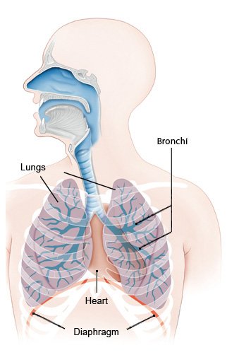
Our lungs are vital organs. They allow us to breathe, ensuring that the oxygen in the air we inhale gets into our blood and reaches all of our body.
The lungs are located in the chest, protected by the ribs in the rib cage. Their structure can be compared to that of an upside-down tree: The windpipe branches into two airways on the left and right, called bronchi, which lead to the lungs. Inside the lungs, the airways keep branching into narrower airways until the air sacs are reached.
A person’s physical fitness greatly depends on how well their lungs and heart work. Your lung function can be measured using various breathing tests.
What happens when you breathe?
When you breathe in (inhale), your chest and lungs expand and air flows into your lungs. When you breathe out (exhale), your lungs get smaller again and air flows out of them. These breathing movements are caused by muscles – particularly the diaphragm and the muscles between the ribs (intercostal muscles). Breathing is controlled by the nervous system, but we normally breathe without thinking about it.
When at rest, adults breathe 14 to 16 times per minute. About half a liter of air is inhaled during one normal breath. When you are more active, your breathing becomes faster and deeper in order to get more oxygen into your blood.
How is oxygen absorbed?
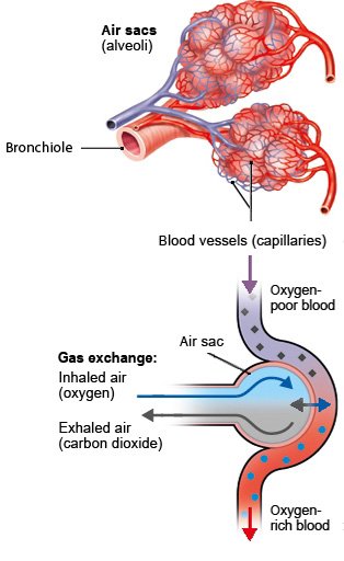
When you breathe in, air containing oxygen enters your windpipe, passes through the bronchi and then reaches the air sacs. These air sacs, called alveoli, look a bit like tiny grapes at the end of the bronchial branches. Healthy lungs have about 300 million air sacs in them. Each air sac is surrounded by a network of fine blood vessels (capillaries).
The oxygen in inhaled air passes across the thin lining of the air sacs and into the blood vessels. This is known as diffusion. The oxygen in the blood is then carried around the body in the bloodstream, reaching every cell. When oxygen passes into the bloodstream from the air sacs, carbon dioxide leaves the blood and passes into the air sacs. Carbon dioxide (CO2) is a waste product of reactions in the cells. You get rid of it when you breathe out. Because these two gases pass between the bloodstream and air sacs in opposite directions, this is referred to as "gas exchange" in the lungs.
Structure of the lower airways
The lower airways are made up of the windpipe (trachea) and the bronchi. In adults, the windpipe is about ten centimeters long. It branches into two main (primary) bronchi, each of which leads to a lung (one on the right side of the body, one on the left). In the lungs, these main bronchi then divide into increasingly smaller bronchi. The smaller ones are known as bronchioles.
The windpipe and bronchi are lined with mucus-producing cells and millions of tiny hair-like projections called cilia. If you breathe in harmful substances like dust or other particles, the mucus and cilia ensure that they don’t stay in your lungs: The foreign particles get caught in the mucus, and the movements of the cilia carry it towards your throat like on a conveyor belt. Once it reaches your throat, you either swallow it or cough it out. As a result, the potentially harmful germs and substances are no longer harmful to you. If larger foreign objects enter the windpipe, a cough reflex is triggered.
Brandes R, Lang F, Schmidt R. Physiologie des Menschen: mit Pathophysiologie. Berlin: Springer; 2019.
Lippert H. Lehrbuch Anatomie. München: Urban und Fischer; 2020.
Pschyrembel Online. 2023.
IQWiG health information is written with the aim of helping people understand the advantages and disadvantages of the main treatment options and health care services.
Because IQWiG is a German institute, some of the information provided here is specific to the German health care system. The suitability of any of the described options in an individual case can be determined by talking to a doctor. informedhealth.org can provide support for talks with doctors and other medical professionals, but cannot replace them. We do not offer individual consultations.
Our information is based on the results of good-quality studies. It is written by a team of health care professionals, scientists and editors, and reviewed by external experts. You can find a detailed description of how our health information is produced and updated in our methods.
Stay informed
Subscribe to our newsletter or newsfeed. You can find our growing collection of films on YouTube.


