How does the spine work?

The spine is our body’s central support structure. It keeps us upright and connects different parts of our skeleton to each other, including the head, ribcage, pelvis, shoulders, arms and legs. Although the spine is made up of a chain of bones, it is flexible. With the help of elastic spinal discs, muscles and ligaments, it can move the body in nearly any direction.

The parts of the spine

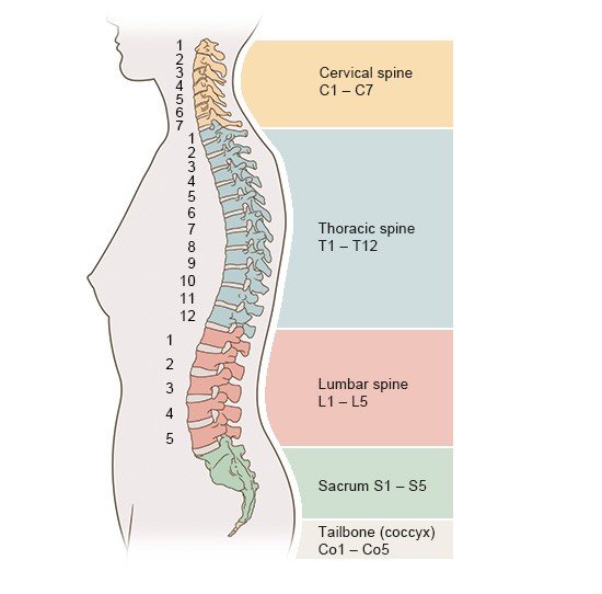
The individual bones in the spine are called vertebrae. Adults typically have 33 vertebrae. They are divided into five areas (from top to bottom):

  • Cervical spine (7 vertebrae)
  • Thoracic spine (12 vertebrae)
  • Lumbar spine (5 vertebrae)
  • Sacrum (5 fused vertebrae)
  • Tailbone, or coccyx (4 fused vertebrae)
This illustration shows a side view of the five parts of an adult spine

Doctors have numbered the vertebrae in each region from top to bottom together with one or two letters standing for each part: Cervical vertebrae are given the letter "C," thoracic vertebrae the letters "Th," and lumbar vertebrae the letter "L," for example. Each region is then numbered from top to bottom, e.g. with C1 to C7 for the cervical vertebrae and so on.

Some regions of the spine are more flexible than others. The cervical spine is the most flexible. It allows us to turn and tilt our head in nearly every direction. The sacrum and tailbone can't be moved, and are made up of only bone in adults. Children still have separate vertebrae making up the sacrum and tailbone, which then gradually grow together.

The lower down the vertebrae are in the spine, the more weight they have to carry. For that reason, the lumbar vertebrae are larger and more stable than vertebrae near the top of the spinal column, for instance. Because of this greater load, normal wear and tear commonly develops here as we age.

What does a healthy spine look like?

Viewed from the side, there are four slight natural curves in a healthy adult spine:

  • The cervical (neck) and lumbar (lower back) sections of the spine curve inward a little. This normal curvature is known as lordosis – cervical lordosis in the neck area, and lumbar lordosis in the lower back.
  • The thoracic (upper back) and sacral (bottom of the spine) sections naturally curve outward a little. An excessive forward-rounding of the spine is known as kyphosis – thoracic kyphosis in the upper back, and sacral kyphosis in the pelvic area.

The natural S-shaped curvature of the spine makes it stable: It helps you keep your balance when you're in an upright position, acts like a shock absorber when you walk, and protects the individual bones in the spine (the vertebrae) from fractures. This curvature develops after birth once more strain is put on the spine as babies start to lift their heads up, crawl and (finally) walk.

In certain diseases, such as Scheuermann's disease (Scheuermann's kyphosis) and ankylosing spondylitis, the curvature of the spine is too strong. This can lead to a poor posture and other problems.

The spine looks straight when viewed from the back. Curvature to the left or right may be a sign of scoliosis, either in children or in old age.

What do vertebrae look like?

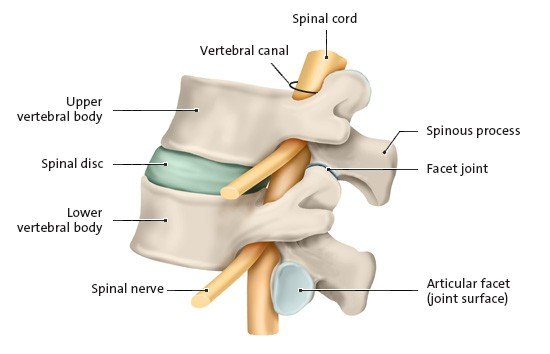
Aside from the first two cervical vertebrae at the top of the spine and the sacrum and tailbone at the bottom end of the spine, all vertebrae have a vertebral body that faces inwards relative to the body and a spinous process that faces outwards. Spinous processes of the vertebrae are visible in some thin people, especially when they bend forwards.

This anatomical illustration shows a side view of two lumbar vertebrae with an intervertebral spinal disc
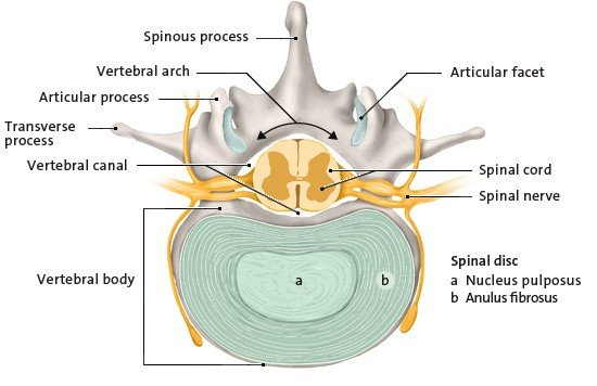
This anatomical illustration shows a cross-section view of a lumbar vertebra with a spinal disc

The other parts of a verebra include:

  • The vertebral arch: It connects the vertebral body to the spinous process, and surrounds an opening called the vertebral foramen. The column formed by the verebral formens stacked in a line is the vertebral canal, which contains the spinal cord.
  • The articular processes: Together with the articular process from the vertebra above, it forms a facet joint.
  • The articular facets: These surfaces are covered with a thin layer of cartilage to allow smooth movement.
  • The transverse processes: They are found on the two sides of the vertebra, and are attached to muscles and ligaments.

What are intervertebral spinal discs for?

There are a total of 23 elastic spinal discs spread out between the vertebrae – except for between the skull and the first cervical vertebra, and between the first and second cervical vertebrae. Each spinal disc consists of a soft gel-like core (nucleus pulposus) that is surrounded by a tough, multi-layered case made of fibrous cartilage (annulus fibrosus).

Spinal discs act as shock absorbers between the vertebrae in our spine. They also act as shock-absorbers when we run or jump, for instance. They keep the spine flexible so that we can lean over and rotate our upper body. Together with the muscles in the upper body, the spinal discs also help to keep the spine stable in order to prevent the vertebrae from slipping.

The spinal discs act a bit like sponges: When we put pressure on our spine, the spinal discs release fluid and become thinner (“compress”); when the pressure is relieved, they absorb fluid again and become thicker (“decompress”). Because we usually put more pressure on our spine during the day and relieve the pressure at night, we are around 1.5 to 2 centimeters shorter (about 0.6 to 0.8 inches shorter) by the end of the day.

Signs of wear and tear may start appearing after many years. As we get older, the spinal discs become narrower, the vertebrae move closer to each other, and the spine becomes more curved. Because of this, older people are usually a few centimeters shorter than they used to be when they were younger.

Wear and tear on the vertebrae and spinal discs (degeneration of the spine) is also known as spondylosis. Many people have degenerative changes in their spine but they don't always have back pain or other symptoms. So when people have these symptoms, it usually isn’t absolutely clear whether they are caused by degeneration of the spine.

Spinal cord and nerves

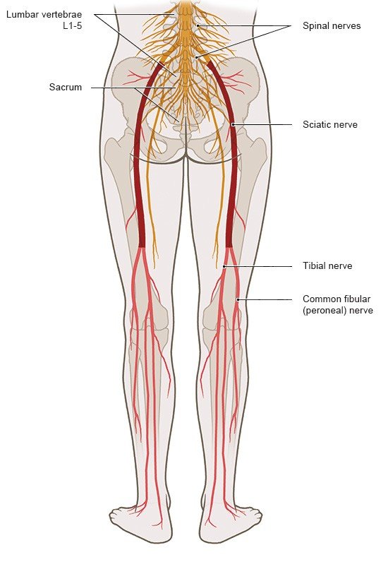
The bones of the spine protect the spinal cord, which runs along the spinal canal. The vertebral arches are slightly indented at the top and bottom of each side. At the point where two vertebrae come together, they form two gaps where the indented parts meet – one on the left and one on the right side of the spine. The spinal nerves connect to one sciatic nerve on the left and on the right in the pelvis. The sciatic nerves run down the back of the legs.

This illustration shows where the sciatic nerve starts and how it runs from the lumbar spine down to the foot.

The spinal nerves carry electrical signals along the spinal cord, from the brain to muscles of the skeleton and internal organs. They also carry sensory information along the spinal cord in the other direction – from the skin, muscles, joints and internal organs to the brain. This sensory information includes things like touch, pressure, cold, warmth and pain. Together, the spinal cord and the brain make up the central nervous system.

The spinal cord ends at the top portion of the . From that point, only a bundle of nerves called the cauda equina continues downward.

Brinjikji W, Luetmer PH, Comstock B et al. Systematic literature review of imaging features of spinal degeneration in asymptomatic populations. AJNR Am J Neuroradiol 2015; 36(4): 811-816.

Deutsche Gesellschaft für Orthopädie und Orthopädische Chirurgie (DGOOC), Deutsche Gesellschaft für Orthopädie und Unfallchirurgie (DGOU), Deutsche Gesellschaft für Neurochirurgie (DGNC) et al. S2k-Leitlinie Konservative, operative und rehabilitative Versorgung bei Bandscheibenvorfällen mit radikulärer Symptomatik (under revision). AWMF register no.: 187-057. 2020.

Jensen RK, Jensen TS, Koes B et al. Prevalence of lumbar spinal stenosis in general and clinical populations: a systematic review and meta-analysis. Eur Spine J 2020; 29(9): 2143-2163.

Klein C. Orthopädie für Patienten: Medizin verstehen. Wirbelsäule, Halswirbelsäule, Brustwirbelsäule, Brustkorb, Lendenwirbelsäule, Schulter, Ellenbogen, Hand, Hüfte, Knie, Fuß. Remagen: Michels-Klein; 2014.

Lippert H, Herbold D, Lippert-Burmester W. Anatomie, Text und Atlas. Munich: Urban und Fischer; 2020.

Menche N. Biologie Anatomie Physiologie. Munich: Urban und Fischer; 2023.

Pschyrembel Online. Wirbelsäule. 2025.

IQWiG health information is written with the aim of helping people understand the advantages and disadvantages of the main treatment options and health care services.

Because IQWiG is a German institute, some of the information provided here is specific to the German health care system. The suitability of any of the described options in an individual case can be determined by talking to a doctor. informedhealth.org can provide support for talks with doctors and other medical professionals, but cannot replace them. We do not offer individual consultations.

Our information is based on the results of good-quality studies. It is written by a team of health care professionals, scientists and editors, and reviewed by external experts. You can find a detailed description of how our health information is produced and updated in our methods.
Comment on this page

What would you like to share with us?

We welcome any feedback and ideas - either via our form or by gi-kontakt@iqwig.de. We will review, but not publish, your ratings and comments. Your information will of course be treated confidentially. Fields marked with an asterisk (*) are required fields.

Please note that we do not provide individual advice on matters of health. You can read about where to find help and support in Germany in our information “How can I find self-help groups and information centers?

Über diese Seite

Updated on August 28, 2025

Next planned update: 2028

Publisher:

Institute for Quality and Efficiency in Health Care (IQWiG, Germany)

Stay informed

Subscribe to our newsletter or newsfeed. You can find our growing collection of films on YouTube.