What happens during an electroencephalogram (EEG)?

Many organs produce electrical signals that can be detected and measured using various techniques. One well-known example is the electrocardiogram (ECG or EKG), which is used to see the electrical currents in the heart. The nerve cells in the brain also produce characteristic electrical currents. Like in an ECG, these currents can be picked up using electrodes on the body’s surface. This kind of measurement is called an electroencephalogram (EEG). In an EEG, the electrodes are placed on specific points on your head and connected to an EEG machine with cables. The electrodes measure the activity of the brain and display it as a graph on a screen.

When is an EEG used?

One reason doctors use an EEG is if they think someone has a neurological (nerve-related) disorder such as epilepsy or brain damage. Sometimes it is used during surgery to monitor the effects of an anesthetic. An EEG can also provide information about how the brain is functioning in patients who are in intensive care or doing a sleep study. It can confirm brain death as well.

EEGs used to play an important role in diagnosing strokes or detecting brain tumors. But nowadays imaging techniques such as CT or MRI scans are used instead.
What does the procedure involve?

A total of 21 electrodes are typically used in an EEG. To make it easier to put the electrodes in the right place on your head, the electrodes and cables are usually attached to a cap that is pulled over your head. Before the test, the electrodes are coated in a special gel that makes it easier to measure the electrical activity.

There is no need to shave your head. Your hair should be clean and free of residues from products such as styling foam, gel or hair spray.

During the measurement, you lie or sit in a relaxed position and stay as still as possible. An assistant will give you directions, such as to open your eyes or to take deep breaths. Sometimes, certain stimuli (such as a flickering light) may be used to trigger brain activity. The recording takes about 20 to 30 minutes.

What can you see on an EEG recording?

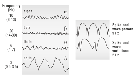
Unlike an ECG, which has a pattern with spikes in it, an EEG shows multiple waves. The shape of these waves mainly depends on how active the brain currently is. This is different when you’re awake or asleep, or when you’re alert or tired. Each wave provides information about the activity of the nerve cells in one specific area of the brain.

Brain waves are divided into the following main types:

  • Alpha waves (frequency 8 to 13 cycles per second (Hz, hertz)): These describe the activity of the brain when you're resting with your eyes closed but you’re awake. This is your baseline.
  • Beta waves (14 to 30 Hz): When your eyes are open, your senses are stimulated and you’re mentally active, these higher, irregular frequencies are recorded.
  • Gamma waves (over 30 Hz): These occur when you're very alert and when you're learning something.
  • Theta waves (4 to 7 Hz): Lower frequencies arise when you are falling asleep or very exhausted, for example.
  • Delta waves (0.5 to 3.5 Hz): The slowest, usually synchronized waves can be observed during deep sleep.


Normal EEG waves (left) and the spike-and-wave pattern in epilepsy (right)
Illustration: Normal EEG waves (left) and the spike-and-wave pattern in epilepsy (right)

Illustration: Normal EEG waves (left) and the spike-and-wave pattern in epilepsy (right)

Everybody’s baseline EEG pattern is different. This means that “normal” EEG recordings can vary greatly from person to person. This variation is even greater in children, and their wave patterns are much slower and less regular than in adults.

If an EEG is done for diagnostic purposes, the focus is on how often waves occur (frequency) and how high they are (amplitude). Deviations from typical wave patterns can be signs of an illness or disorder. During epileptic seizures, for example, some of the waves are particularly high and steep (spike-and-wave pattern). Many medications that affect the brain also change the EEG pattern.

Hacke W (Ed). Neurologie. Berlin: Springer; 2016.

Schmidt RF, Lang F, Heckmann M (Ed). Physiologie des Menschen. Berlin: Springer; 2011.

IQWiG health information is written with the aim of helping people understand the advantages and disadvantages of the main treatment options and health care services.

Because IQWiG is a German institute, some of the information provided here is specific to the German health care system. The suitability of any of the described options in an individual case can be determined by talking to a doctor. informedhealth.org can provide support for talks with doctors and other medical professionals, but cannot replace them. We do not offer individual consultations.

Our information is based on the results of good-quality studies. It is written by a team of health care professionals, scientists and editors, and reviewed by external experts. You can find a detailed description of how our health information is produced and updated in our methods.

Comment on this page

What would you like to share with us?

We welcome any feedback and ideas - either via our form or by gi-kontakt@iqwig.de. We will review, but not publish, your ratings and comments. Your information will of course be treated confidentially. Fields marked with an asterisk (*) are required fields.

Please note that we do not provide individual advice on matters of health. You can read about where to find help and support in Germany in our information “How can I find self-help groups and information centers?

Über diese Seite

Updated on January 11, 2023

Next planned update: 2026

Publisher:

Institute for Quality and Efficiency in Health Care (IQWiG, Germany)

How we keep you informed

Follow us on Twitter or subscribe to our newsletter or newsfeed. You can find all of our films online on YouTube.