How does mechanical ventilation work during an operation?

People who undergo an operation under general anesthetic don’t notice what's going on around them in the operating room. They aren't aware of mechanical ventilation used during the procedure, either. Which types of mechanical ventilation can be used during surgery, and what exactly do they involve?

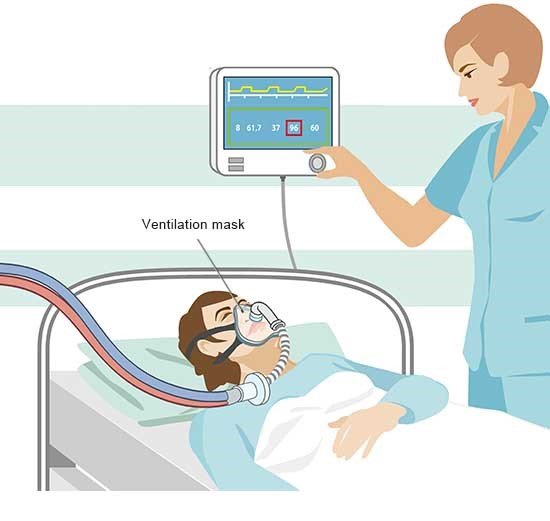
Why is mechanical ventilation even needed during an operation?

An anesthetic is necessary to make sure that a surgical procedure doesn't cause any pain. It is sometimes enough to just anesthetize (numb) one part of the body, such as the edges of a wound, only one arm, or the lower abdomen. However, a general anesthetic is usually needed for more extensive surgery. It deactivates our consciousness and the pain receptors in our body. Depending on how strong the anesthetic is, it can also stop our breathing. A ventilator then has to take over our breathing.

How do ventilation masks work?

A mask is placed on your face which firmly covers the nose and mouth. A ventilator can be attached to the mask to constantly provide fresh, oxygen-rich air to the airways.

A manual resuscitator or bag-valve-mask (BVM) can be attached to the mask for a short period or in emergencies. The anesthetist pumps it rhythmically to push the air from the bag into the airways. Valves make sure that every time they let go, the exhaled air escapes and the bag fills back up with fresh air.

Doctors also refer to mask ventilation as “non-invasive” ventilation.

Illustration: Mask ventilation

What is the difference between a laryngeal mask airway and a ventilation mask?

Laryngeal mask airways (LMAs) are now used for a lot of procedures. The mask resembles an ear with two tubes attached. It is not placed over your mouth and nose, but rather inserted into the throat above the larynx. To do so, the doctor has to feed the mask in through your mouth. Once the right position is found, a little air is fed into the inflatable edge of the mask through the thinner tube. It then covers the larynx better. The ventilation device or the manual resuscitator is then connected to the thicker tube.

Because the laryngeal mask airway is deployed deep inside the throat, doctors also refer to it as “invasive ventilation.” It is only introduced once the anesthetic has started to work, so the person being operated on doesn't notice anything.

Illustration: Laryngeal mask airway ventilation

What is mechanical ventilation via intubation?

Intubation usually refers to when the anesthetist introduces a flexible plastic tube (called a tracheal or endotracheal tube) into the windpipe. A ventilator is then connected to the other end of the tube. A manual resuscitator can be used instead as a temporary solution.

Intubation is also referred to as invasive ventilation. As when the laryngeal mask airway is inserted, the person being operated on doesn't feel the tube being put in because the anesthetic is already taking effect.

Illustration: Air is fed directly into the windpipe via the tube

How do ventilators work?

Ventilators regularly pump air into the lungs via tubes. The anesthetist can control exactly what should happen during ventilation, for instance:

  • how long each breath lasts,
  • how much air flows into the lungs,
  • the pressure in the airways, or
  • the oxygen concentration of the air.

Anesthetic gases and moisture can also be mixed in with the ventilating air.

The physician can keep an eye on all key measurements using a monitor and then adjust the device when needed.

Illustration: Ventilator in a hospital

What are the possible side effects and complications?

One complication is that the contents of the stomach can flow back up the food pipe into the mouth and throat and then enter the windpipe. That is called aspiration. Patients need to have an empty stomach before an operation to minimize the risk of that happening.

This risk is even lower when intubation is used compared to a ventilation mask or a laryngeal mask airway, because the end of the tube that is introduced into the windpipe is surrounded by an inflatable plastic cuff. As soon as it has been inserted far enough, the cuff is filled with a little air. That fixes the tube in place and seals off the windpipe. This makes it impossible for the contents of the stomach to pass by the tube and enter the windpipe. After surgery, the air has to be let out of the cuff before the tube is removed.

More medication is needed during intubation than for mask ventilation though, which can affect circulation. Injuries during intubation and hoarseness and irritations of the throat are also possible following ventilation. Complications can also occur if the tube is not inserted correctly into the windpipe. For example, it might be inserted into the adjacent opening of the food pipe or be pushed too far down into the lungs. That is why doctors need good visibility when inserting the tube. A hook-shaped instrument with a light on it called a laryngoscope can be helpful. It keeps the airways open while also illuminating the throat. The doctors can then check that the tube is actually going into the windpipe. They also listen to the lungs and analyze the exhaled air after intubation as an additional check that the tube has been positioned correctly.

Side effects of ventilation during an operation such as damage to the lungs or circulatory system or infections with hospital germs are rare, and are more of an issue with longer term mechanical ventilation or if the lungs are already damaged.

When are the different types of ventilation considered?

A number of factors dictate which type of ventilation is used when:

  • the type of operation,
  • the type of anesthetic needed, and
  • the condition of the person being operated on.

Individual risk of complications is the most important factor. If a fast operation is needed, there is not enough time to wait for the person's stomach to be empty. Intubation is then usually used to minimize the risk of aspiration. Ventilation and laryngeal masks are also an option for scheduled procedures. Intubation is the general rule for longer, more complicated operations, even if the patient has an empty stomach.

It is also possible that the insertion of a tube is difficult or unsuccessful. The doctors then have to consider: if mask ventilation can be used instead, whether a different type of anesthetic is possible, or if the procedure can be postponed.

When else is mechanical ventilation needed?

Even without general anesthetic, you can also fall into a deep unconscious state where you can no longer breathe unaided, for instance due to severe head injuries or significant blood loss. Experienced first aiders can use mouth-to-mouth resuscitation in those types of emergency situations to make sure the unconscious person is still getting enough oxygen. As soon as experts take over, ventilation can be continued using a bag-valve-mask or ventilator.

Lung problems are often the reason people have to be mechanically ventilated. If they are significantly impaired, for instance due to severe pneumonia, normal breathing will no longer be enough to provide the body with sufficient oxygen. Ventilation therapy is helpful then.

Deutsche Gesellschaft für Anästhesiologie und Intensivmedizin (DGAI). S1-Leitlinie Atemwegsmanagement. AWMF-Registenr.: 001-028. 2015.

Deutsche Gesellschaft für Anästhesiologie und Intensivmedizin (DGAI). S3-Leitlinie Invasive Beatmung und Einsatz extrakorporaler Verfahren bei akuter respiratorischer Insuffizienz. AWMF-Registernr.: 001-021. 2017.

Deutsche Gesellschaft für Pneumologie und Beatmungsmedizin (DGP). Prolongiertes Weaning (S2k-Leitlinie). AWMF-Registernr.: 020-015. 2019.

Deutsche Gesellschaft für Pneumologie und Beatmungsmedizin (DGP). S2k-Leitlinie Nichtinvasive und invasive Beatmung als Therapie der chronischen respiratorischen Insuffizienz (Revision 2017). 2017.

Deutsche Gesellschaft für Pneumologie und Beatmungsmedizin (DGP). S3-Leitlinie Nichtinvasive Beatmung als Therapie der akuten respiratorischen Insuffizienz. AWMF-Registernr.: 020-004. 2015.

Pschyrembel Online. 2022.

Striebel HW. Anästhesie, Intensivmedizin, Notfallmedizin. Stuttgart: Thieme; 2020.

IQWiG health information is written with the aim of helping people understand the advantages and disadvantages of the main treatment options and health care services.

Because IQWiG is a German institute, some of the information provided here is specific to the German health care system. The suitability of any of the described options in an individual case can be determined by talking to a doctor. informedhealth.org can provide support for talks with doctors and other medical professionals, but cannot replace them. We do not offer individual consultations.

Our information is based on the results of good-quality studies. It is written by a team of health care professionals, scientists and editors, and reviewed by external experts. You can find a detailed description of how our health information is produced and updated in our methods.

Comment on this page

What would you like to share with us?

We welcome any feedback and ideas - either via our form or by gi-kontakt@iqwig.de. We will review, but not publish, your ratings and comments. Your information will of course be treated confidentially. Fields marked with an asterisk (*) are required fields.

Please note that we do not provide individual advice on matters of health. You can read about where to find help and support in Germany in our information “How can I find self-help groups and information centers?

Über diese Seite

Created on May 2, 2022

Next planned update: 2025

Publisher:

Institute for Quality and Efficiency in Health Care (IQWiG, Germany)

How we keep you informed

Follow us on Twitter or subscribe to our newsletter or newsfeed. You can find all of our films online on YouTube.