What kinds of eye examinations are there?

Eye examinations tend to follow a certain pattern: After talking with your doctor about the type and severity of your symptoms, both eyes will usually be tested to measure the visual acuity (sharpness of sight). Then the doctor will look at your eyes from the outside, take a closer look at them using a slit lamp, and measure the (the pressure inside the eyeball). Eye drops are given before some tests to dilate (enlarge) the of the eye for a certain amount of time. This makes it easier for the doctor to see the back of the eye. Depending on the symptoms and any other medical conditions you may have, the eye examination might be carried out differently and additional tests might be done.

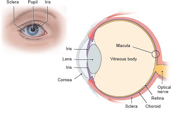
Illustration: Structure of the eye

What do general eye examinations involve? 

Eye examinations may be necessary for various reasons, for instance due to external eye injuries or sudden vision problems. Vision also tends to get worse with age, so many people end up going to see an eye doctor at some point.

General eye examinations typically involve testing your vision and your pupillary response (how your pupils react). Pupils usually automatically contract and get smaller when light enters the eye.

The doctor first looks at the eye from the outside to see whether, for instance, the conjunctiva or eyelids are red and inflamed. The conjunctiva are the membranes lining the exposed part of the eyeball and inner surface of the eyelids. If your eye hurts, it is important to describe exactly where it hurts – e.g. on the surface of the eye, inside the eye or only when you move your eye? Eye pain can also radiate to your forehead or temples.

How is visual acuity measured?

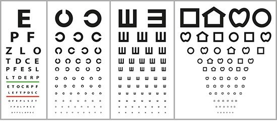
If you aren't sure whether your vision has got worse, you can have your visual acuity (sharpness of sight) tested by an eye specialist. To do this eye test, one of your eyes is covered and you are asked to look at an eye chart with your other eye. The chart may have letters, numbers or C-shapes on it. These get smaller as you go down the chart.

Illustration: Various types of eye charts

Your visual acuity is measured by determining which of the figures you can still recognize from five meters away. The eye doctor will also check your near vision by holding a card with a similar chart on it at a distance of about 30 centimeters (12 inches). If necessary, this eye test can then be repeated using corrective lenses.

Technically speaking, visual acuity is a measure of how well you can see two points as being separate. The closer together the points are and the further away they are from you, the more difficult this becomes.

How else can your vision be tested?

Vision is often assessed using other tests too. These include color vision tests, where you are asked to identify letters or numbers in multicolored pictures. Eye movement and the position of your eyes relative to one another are important too. In children who have a squint, for example, the relative position of their eyes is abnormal. Tests to check your field of vision are also common.

What are visual field tests?

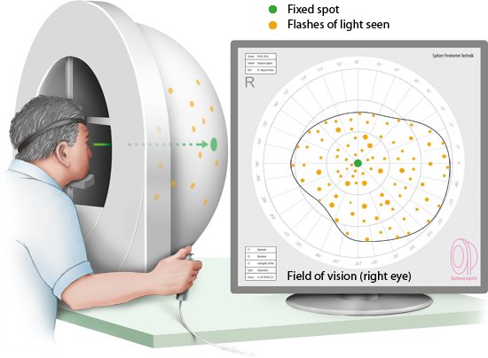
Your field of vision (or visual field) is what you can see without moving your eye. The visual field test is done using a bowl-shaped instrument known as a perimeter. With your head supported by a chin rest, you focus on a fixed spot at the center of the bowl. Lights flash in the bowl, and may move around or become increasingly brighter. If you see a flash or notice a change, you press a button. This makes it possible to measure the borders of the areas in which you can still perceive something. The more areas in your field of vision are tested, the more accurate the test is – but it will then take longer as a result. Although visual field tests aren't painful, they can be quite tiring because you have to concentrate for a long time.

Illustration: Visual field test: Press a button everytime you see a flash

Your field of vision is very important as it helps you know what is around you. Because various medical conditions affect the field of vision, these tests are often done if damage to the or optic nerve is suspected, or if someone has headaches or dizziness for no known reason. Eye diseases like glaucoma and age-related macular degeneration also lead to changes in the field of vision.

What are slit-lamp examinations?

A slit lamp is a microscope that allows the doctor to look inside of the eye from the outside. It is one of the main instruments used by eye specialists. You are asked to place your head on a chin rest. The doctor sits across from you and shines the light into your eyes, one at a time.

As its name suggests, the lamp projects a narrow, slit-shaped beam of light into the eye. Depending on how the lamp is set, it is possible for the doctor to see nearly all parts of the eye magnified up to 60 times: the front, middle and back of the eye, and even as far back as the in the fundus of the eye. Modern slit lamps also include a device that measures (the pressure inside the eyeball).

What is ophthalmoscopy?

A special device known as an ophthalmoscope can be used to look at the fundus (back) of the eye through the and examine structures like the , optic nerve, blood vessels and the choroid coat. This examination is known as ophthalmoscopy or funduscopy ("fundus" is the Latin word for "bottom" or "base"). About 20 minutes before the examination, the pupils of the eyes are dilated (enlarged) using eye drops.

In the direct exam, the doctor positions the ophthalmoscope close to the eye and shines a beam of light directly into it to see a small magnified section of the back of the eye. This is particularly useful for examining the structures at the center of the eye's fundus, including the optic nerve, the blood vessels and the .

In the indirect exam, the doctor uses a condensing lens with a bright light to examine the eye from a distance of about 60 centimeters. This allows them to see a larger area of the fundus, providing a better overview as well as a three-dimensional image. A slit lamp is sometimes used at the same time. Doing so greatly magnifies the and provides better lighting.

Conditions such as retinal detachment and optic nerve damage can be diagnosed using ophthalmoscopy. It can also be a good idea to examine the small blood vessels in the eye in people who have diabetes or high blood pressure because both of these conditions can damage blood vessels in the eye.

If your pupils are dilated using eye drops, your eyes will be very sensitive to light and your vision will be blurred for a few hours after the examination. For this reason, you are only allowed to drive a car or operate machinery again once the effect of the eye drops has worn off.

How is intraocular pressure measured?

The measurement of (tonometry) is one of the most common eye examinations. A fluid called aqueous humor is constantly being produced inside the eye. The eye's results from the balance between how much new aqueous humor is being produced and how much of it is flowing out of the eye. The pressure increases if the aqueous humor can't drain properly and starts to build up. An of between 10 and 21 mmHg (millimeters of mercury) is considered to be normal in adults.

Various instruments can be used to measure the pressure in the eye. In what is known as applanation tonometry, the is first numbed using eye drops. Then a small measuring device is placed against the surface of the , putting a little pressure on the eye.

The device measures how much pressure is needed to flatten the by 3 millimeters. This is a measure of the pressure inside the eye.

Intraocular pressure can also be measured using an approach known as non-contact tonometry. Here the pressure is measured by applying a short puff of air to the . Neither of these examinations are painful.

Further eye examinations

Other eye examinations can be done to confirm certain diagnoses and help plan treatment. For instance, the can be examined using fluorescein angiography. This involves injecting a fluorescent dye into a vein in the arm. The dye spreads through all the blood vessels in the body, including those in the . The doctor can then use a special camera to detect changes in the blood vessels of the eye.

Optical coherence tomography (OCT) is another type of eye examination. OCT uses a laser to measure things like the thickness of the . This technique can be applied to detect the build-up of fluid in the .

Andreae S, Avelini P, Berg M, Blank I, Burk A. Lexikon der Krankheiten und Untersuchungen. Stuttgart: Thieme; 2008.

Dahlmann C. Basics Augenheilkunde. München: Urban und Fischer; 2020.

Pschyrembel Online. 2022.

IQWiG health information is written with the aim of helping people understand the advantages and disadvantages of the main treatment options and health care services.

Because IQWiG is a German institute, some of the information provided here is specific to the German health care system. The suitability of any of the described options in an individual case can be determined by talking to a doctor. informedhealth.org can provide support for talks with doctors and other medical professionals, but cannot replace them. We do not offer individual consultations.

Our information is based on the results of good-quality studies. It is written by a team of health care professionals, scientists and editors, and reviewed by external experts. You can find a detailed description of how our health information is produced and updated in our methods.
Comment on this page

What would you like to share with us?

We welcome any feedback and ideas - either via our form or by gi-kontakt@iqwig.de. We will review, but not publish, your ratings and comments. Your information will of course be treated confidentially. Fields marked with an asterisk (*) are required fields.

Please note that we do not provide individual advice on matters of health. You can read about where to find help and support in Germany in our information “How can I find self-help groups and information centers?

Über diese Seite

Updated on November 23, 2022

Next planned update: 2025

Publisher:

Institute for Quality and Efficiency in Health Care (IQWiG, Germany)

How we keep you informed

Follow us on Twitter or subscribe to our newsletter or newsfeed. You can find all of our films online on YouTube.