What happens during cardiac catheterization?

Cardiac catheterization is a procedure used to show on a computer monitor diseases that affect the heart. A cardiac is a thin, flexible tube. During catheterization, x-ray monitoring is used to guide the through a blood vessel from the groin or arm to the heart.

This procedure is often done to make the coronary arteries visible. The medical term for this is coronary angiography. Any narrowed blood vessels can be treated right away using the cardiac .

The may need to be advanced to the heart for other diseases – such as heart valve problems or an irregular heartbeat (cardiac arrhythmia). Then doctors are often able to treat the heart directly by using the to put in a new heart valve or to carry out cardiac ablation, for example.

What different types of cardiac catheterization are there?

There are two main types of cardiac catheterization:

  • Right heart catheterization (venous or flow-directed ): The is inserted through a vein in the groin region or at the elbow and then advanced to the right ventricle and the pulmonary artery. Flow-directed catheters have a small inflatable balloon at the tip, allowing them to be carried by the flow of blood. The is used to measure the pressure levels in the right ventricle and the pulmonary artery. A contrast medium is injected through the so that the right chamber, the circulation of blood and the function of the cardiac valves can be examined.
  • Left heart catheterization (arterial ): This is advanced through an artery in your groin region, elbow or wrist and into the heart’s left ventricle. After a contrast medium has been injected it’s possible to examine the left ventricle, the aorta, the circulation of blood and the performance of the cardiac valves.
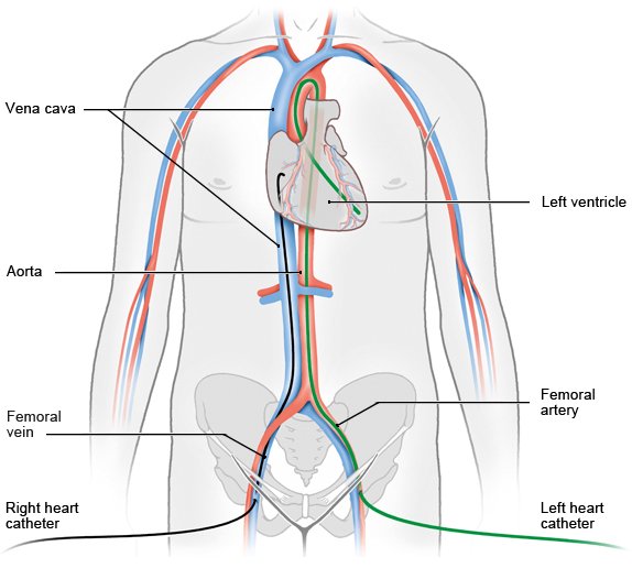
Illustration: Right and left heart catheterization, depicting the catheter reaching the heart with respective entry points

What is coronary angiography?

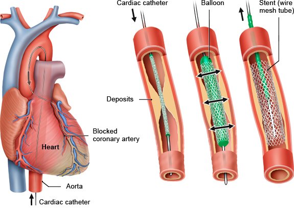
Coronary angiography (an x-ray examination of the coronary arteries) is also possible with the commonly used left-heart catheterization procedure. Contrast medium is injected into the coronary arteries to find possible constrictions. Treatment can also be performed during the examination: If narrowed arteries (coronary artery disease, CAD) are found, they can be pushed open using a balloon affixed to the tip of the . The can also be used to implant a metal tissue support (stent) to keep the blood vessel open.

Illustration: Cardiac catheter with balloon and stent

But coronary angiography (left heart catheterization) can't always find out whether a narrowed artery needs to be expanded. Measurements can then be done to see how affected the blood flow is in the narrowed section. A flexible wire with a built-in sensor is pushed through the into the narrowed artery. The sensor measures the pressure before and after the narrowed section: The bigger the pressure difference is, the more affected the blood flow towards the heart is. The measured value is called the myocardial fractional flow reserve (FFR).

What else can be examined using a catheter?

Tissue samples can also be taken during right heart and left heart catheterization. Doctors can also check possible causes of arrhythmia. Specialized catheters may be used to measure how much blood the heart pumps in one minute. This can help doctors find out whether the heart is performing poorly.

When is cardiac catheterization used?

Cardiac catheterization is especially well-suited for the following situations:

  • Symptoms such as chest pain or tightness and shortness of breath, or after a heart attack. Left heart catheterization can help to determine the location and severity of a possible narrowed coronary artery.
  • Coronary artery disease: A narrowed or blocked blood vessel can be expanded with left heart catheterization. Blood flow at the narrowed section can be measured beforehand (FFR value).
  • Suspected degenerative changes or dysfunction affecting the cardiac valves.
  • Before heart surgery: If heart valve or surgery is planned, the heart muscle tissue and the coronary arteries are checked for any abnormal changes using cardiac catheterization.
  • For biopsies: Heart muscle tissue can be taken using a in case of myocarditis (heart muscle ).
  • For an irregular heartbeat (cardiac arrhythmia).

What needs to be done to prepare?

Before starting the doctor first needs to know about any other medical problems you may have, such as diabetes, infections, bleeding disorders or allergies. It is also important to let the doctors know what medication you use (like anticoagulants, beta blockers or antidiabetics). A number of tests need to be completed before the procedure can be started:

  • ECG
  • Blood pressure measurement
  • Blood testing: This serves to find out your blood count, and determine other parameters describing blood clotting ability and kidney and thyroid function. The latter is useful if a contrast medium containing iodine is injected.

Additional examinations ate sometimes done, possibly including a lung x-ray or an ultrasound of the heart (echocardiography).

What happens during the examination?

The procedure is done in a catheterization laboratory (sometimes called a “cath lab”). Often the lab is in a hospital, but catheterization can also be done in an outpatient setting at a specialist clinic. You’re not allowed to eat anything for about six hours before the examination. The area of skin right at the insertion site in your groin region or on your arm may need to be shaved right before the procedure. You can be given a local anesthetic if you like.

The doctor punctures a blood vessel and then inserts the to move it up to your heart. While this is done, the situation is monitored on a computer screen. Usually you don’t feel anything because the inside walls of the blood vessels aren’t sensitive to pain.

An x-ray contrast medium is injected through the into the chambers of the heart and the coronary arteries to make them visible on the computer screen. This may feel warm for a short time. You may also feel temporary heart pain if one of the narrowed arteries is pushed open. This usually goes away as soon as the expansion is completed.

You can watch pictures of your own heart on the screen during the examination. The examination takes usually about 30 minutes. But it may also take much longer depending on what the doctors find and whether further measurements, expansion of the blood vessels or stent implantation are needed.

A pressure bandage is applied to the insertion site afterwards to prevent bleeding. Doctors will ask you to stay at the hospital another five hours for your own safety. You’ll need to continue to stay in a lying position for about four hours if the was inserted in the groin region. And an additional two days of monitoring is usually required if a narrowed artery was detected and treated.

It is always important to rest at home for the first few days and avoid lifting heavy objects.

What risks does cardiac catheterization involve?

Cardiac catheterization is classified as a minimally invasive procedure, because no larger incisions are needed. It's considered to be a safe examination method: only about 1 out of 100 examinations results in complications. But if complications do occur, they can be severe – after all, it is a heart procedure. Because of this, your doctor must talk to you about the risks, possible consequences and alternative methods for examining your heart one day or more in advance of the procedure, unless it’s an emergency. After that you need to provide your written informed consent.

The risks include:

The risk of complications increases in people who already have severe cardiac disease or other related conditions.

Bundesärztekammer (BÄK), Kassenärztliche Bundesvereinigung (KBV), Arbeitsgemeinschaft der Wissenschaftlichen Medizinischen Fachgesellschaften (AWMF). Nationale Versorgungsleitlinie Chronische KHK (S3-Leitlinie, in Überarbeitung). AWMF-Registernr.: nvl-004. 2019.

Deutsche Gesellschaft für Kardiologie – Herz-und Kreislaufforschung (DGK). Leitlinie Diagnostische Herzkatheteruntersuchung. Clin Res Cardiol 2008; 97(8): 475-512.

Erdmann E. Klinische Kardiologie. Krankheiten des Herzens, des Kreislaufs und der herznahen Gefäße. Berlin: Springer; 2011.

Institut für angewandte Qualitätsförderung und Forschung im Gesundheitswesen (AQUA). Ergebnisse der Qualitätssicherung zur Koronarangiographie und Perkutanen Koronarintervention (PCI). In: Qualitätsreport 2013. 2014. S. 61-65.

Institute for Quality and Efficiency in Health Care (IQWiG, Germany). Measurement of myocardial fractional flow reserve (FFR) in coronary heart disease: Final report: Comission D15-02. 14.11.2016. (IQWiG reports; Volume 462).

Stierle U. Klinikleitfaden Kardiologie. München: Urban und Fischer; 2017.

IQWiG health information is written with the aim of helping people understand the advantages and disadvantages of the main treatment options and health care services.

Because IQWiG is a German institute, some of the information provided here is specific to the German health care system. The suitability of any of the described options in an individual case can be determined by talking to a doctor. informedhealth.org can provide support for talks with doctors and other medical professionals, but cannot replace them. We do not offer individual consultations.

Our information is based on the results of good-quality studies. It is written by a team of health care professionals, scientists and editors, and reviewed by external experts. You can find a detailed description of how our health information is produced and updated in our methods.

Comment on this page

What would you like to share with us?

We welcome any feedback and ideas - either via our form or by gi-kontakt@iqwig.de. We will review, but not publish, your ratings and comments. Your information will of course be treated confidentially. Fields marked with an asterisk (*) are required fields.

Please note that we do not provide individual advice on matters of health. You can read about where to find help and support in Germany in our information “How can I find self-help groups and information centers?

Über diese Seite

Updated on June 6, 2023

Next planned update: 2026

Publisher:

Institute for Quality and Efficiency in Health Care (IQWiG, Germany)

Stay informed

Subscribe to our newsletter or newsfeed. You can find our growing collection of films on YouTube.