How do the bones work?

The bones in our bodies have many different jobs to do. They carry our body weight and are exposed to various strains whenever we stand, sit or move around.

Their inner structure makes them strong and at the same time so elastic that they can withstand pressure and torsion, as well as slight bending and rotation. Almost all bones are connected with the flexible skeleton by joints, tendons, ligaments, and muscles.

Bones have an active metabolism which plays an especially important role in the production of blood and in our body's levels.

Bone structure

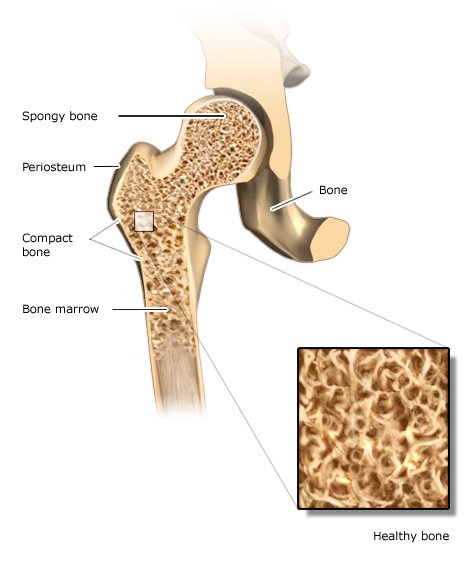
Our bones’ outer walls are referred to as the outer bone layer (compacta). This layer is hard and especially strong. Inside bones there is a supporting structure with interconnecting bony plates and rods called trabeculae. It is called spongy bone because of its sponge-like structure, but is sometimes also referred to as trabecular or cancellous bone. Long bones like the arms and legs also have a bone marrow cavity.

The bones have good blood circulation: Numerous veins pass through them. The spongy bone and the marrow cavities contain red bone marrow, which produces blood cells and what is known as fatty marrow from fat tissue. In childhood, many of our bones contain red bone marrow, but in adulthood red bone marrow is only found in certain bones like the ribs and vertebrae, the sternum (breastbone) and the pelvis.

Illustration: Healthy bone structure

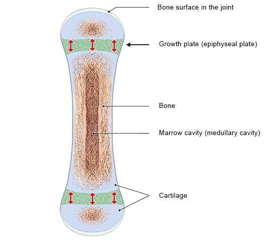
Some bones develop from connective tissue, like the skull. Others start off as cartilage that is later replaced by bone tissue. The arm and leg bones are examples of this. But they do not completely turn into bone for as long as we are still growing: A small amount of cartilage remains at the ends of the bones. These parts are called growth or epiphyseal plates. New cartilage is constantly being produced there, which later becomes bone. That allows the bones to keep growing longer. Towards the end of puberty, the growth plates gradually turn into bone as well, and our bones are fully grown.

Illustration: Long bones with epiphyseal plates

How does bone tissue regenerate?

The mineral content of our bones is crucial for their strength. Calcium phosphate is the most important of these minerals. It makes the bones harder and stronger. The more minerals a bone has in it, the harder and denser it is. But the strength of a bone also depends on the shape and density of the spongy bone inside.

Two kinds of living cells that make our bones a kind of permanent construction site are walled inside the bone tissue: The osteoblasts build layer upon layer of new bone material all around themselves and make the bone grow. The osteoclasts do the opposite: They break down old bone tissue. Together, these two types of cells make sure that every bone can adapt to the strains it is put under and can repair itself after a fracture.

As long as the same amount of tissue is produced as is broken down, the bone gradually regenerates without getting weaker. That means all of the bone tissue in the skeleton is completely replaced about every eight years. Various hormones such as sex, growth and thyroid control this regeneration process.

Adult skeletons weigh about seven to nine kilograms. Bone cells make up about one percent of the bone, the rest is bone tissue. It is made up of 25% water and 25% protein such as . Minerals such as phosphate make up 50% of the bone.

Why are bone fractures nore common in older age?

Bones are still growing in children and teenagers: They grow longer, heavier, and more dense. Our bones are most stable around the age of 30. After that, our bodies slowly start to break down more bone than they produce. That process speeds up at around the age of 50, especially in women. Up until menopause, the female sex hormone estrogen protects the bones by slowing down bone loss. Bone tissue is then broken down more quickly when estrogen levels gradually go down after menopause.

The risk of fractures can increase in older age and osteoporosis can develop. But there are lots of things you can do to protect and strengthen your bones, even in older age. Exercise is most important, because if enough strain is put on the bones their metabolism also remains active and new bone tissue is produced.

What happens when bones and cartilage wear down?

The ends of the bones in our joints are covered in a layer of cartilage. If that cartilage is very worn, perhaps due to old age, the bone also changes. This kind of wear (degenerative changes) of the bones (osteo) and cartilage (chondro) is called osteochondrosis.

If a vertebral disc between two vertebrae gets thinner, bone growths often develop on the edges of the vertebra (osteophytes). That can also happen on worn joints, like on the knee or hip. These growths can be like bulges or bumps. They develop because the body tries to make the joint bigger to spread the pressure across a larger area. Depending on their size, osteophytes can go unnoticed, but they can also cause pain and restrict mobility in the joint. They can cause narrowing (stenoses) of the spinal canal where the bone marrow is found.

What is osteochondrosis dissecans?

Especially in children and teenagers, a fragment of bone and cartilage can become loose under a joint surface because of bone wear. This is called “osteochondrosis dissecans.” If the loose fragment stays where it is, it only causes mild pain or might not even be noticed. But it can also become completely detached and move into the joint. A “joint mouse” like this can cause painful swelling or block the joint. Joint mice are usually broken down over time. But because the area in the joint where the fragment broke off never heals completely, there is an increased risk of osteoarthritis.

Experts think that along with hereditary predisposition, severe joint strain, metabolic disorders, and can also be a risk factor for developing a joint mouse. Osteochondrosis dissecans is most common in the knee joint, but it can affect any joint.

Brandes R, Lang F, Schmidt R (Ed). Physiologie des Menschen: mit Pathophysiologie. Berlin: Springer; 2019.

Klein C. Orthopädie für Patienten. Remagen: Verlag Michels-Klein; 2014.

Vaupel P, Schaible HG, Mutschler E. Anatomie, Physiologie, Pathophysiologie des Menschen. Stuttgart: Wissenschaftliche Verlagsgesellschaft; 2015.

IQWiG health information is written with the aim of helping people understand the advantages and disadvantages of the main treatment options and health care services.

Because IQWiG is a German institute, some of the information provided here is specific to the German health care system. The suitability of any of the described options in an individual case can be determined by talking to a doctor. informedhealth.org can provide support for talks with doctors and other medical professionals, but cannot replace them. We do not offer individual consultations.

Our information is based on the results of good-quality studies. It is written by a team of health care professionals, scientists and editors, and reviewed by external experts. You can find a detailed description of how our health information is produced and updated in our methods.

Comment on this page

What would you like to share with us?

We welcome any feedback and ideas - either via our form or by gi-kontakt@iqwig.de. We will review, but not publish, your ratings and comments. Your information will of course be treated confidentially. Fields marked with an asterisk (*) are required fields.

Please note that we do not provide individual advice on matters of health. You can read about where to find help and support in Germany in our information “How can I find self-help groups and information centers?

Über diese Seite

Updated on January 18, 2024

Next planned update: 2027

Publisher:

Institute for Quality and Efficiency in Health Care (IQWiG, Germany)

How we keep you informed

Follow us on Twitter or subscribe to our newsletter or newsfeed. You can find all of our films online on YouTube.