How do ultrasound examinations work?

Ultrasound is used to be able to see the organs inside the body. It can help doctors to diagnose various medical conditions. Other medical terms for ultrasound are and ultrasonography.

Ultrasonic sound waves pass through tissue in the body without damaging it. Unlike x-ray examinations, ultrasound does not use any radiation.

How does ultrasound work?

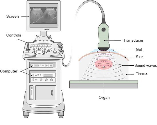
Doctors use an ultrasound machine for the examination. It usually looks like a cabinet with a computer-like device and a screen. A hand-held device called a transducer is connected to it by a cable.

Illustration: Ultrasound machine with a screen and transducer – as described in the information

The transducer has special crystals inside it. When the machine is switched on, the transducer acts like a loudspeaker and produces sound waves that the human ear can't hear. These sound waves pass into the body when the transducer is held against your skin. The sound waves then bounce back from the organs and tissues inside the body, like an echo.

Different types of tissue reflect the sound waves differently. A computer uses these echoes to determine the position, shape and structure of the organs. This is then displayed on the attached monitor as an ultrasound image. The results of the examination and individual images can be saved and printed.

Some ultrasound devices can also determine the direction and speed at which blood is flowing through blood vessels or the heart. That is known as Doppler ultrasound.

What preparations need to be made?

You usually don’t need to do anything to prepare for an ultrasound examination. If you do, your doctor will let you know: For instance, in order to see the urinary bladder better, it’s better if it's at least partially full. So you may be asked to not go to the toilet right before the examination. Air and partially digested food may cause problems with the ultrasound image when the bowel and nearby organs are examined more closely. So you may be asked to come to an ultrasound examination of the abdomen on an empty stomach.

What does the examination involve?

Typically, you will have to lie down on your back. Depending on what part of the body needs to be examined, you may be asked to move into a special position – like turning to your side so that doctors can get better images of your heart or kidneys, for example. When examining the neck, a small cushion is placed under your neck so that your head tilts back a little.

What happens during the examination?

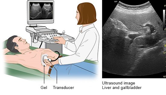
The doctor will put some clear gel on the transducer or the part of your body that will be examined. This gel is used to improve the contact between the transducer and your skin. Then the doctor will press the transducer against your skin and move it from side to side. This usually doesn't hurt, but you may feel a little pressure. You might need to briefly hold your breath during the examination for doctors to record a clearer image.

The time it takes to complete an ultrasound examination can vary quite a bit. It may be over in just a few minutes, but sometimes it takes more than half an hour – depending on which and how many organs are examined.

What parts of the body are examined?

Ultrasound can be used to examine nearly the entire body:

  • The most commonly examined structures in the head and neck area are the thyroid gland and the blood vessels.
  • In the chest area, ultrasound is mostly used to look at the heart (also called an echocardiogram or “echo”). But ultrasound can also be used to look at the edges of the lungs. Closer to the center of the body, the air in the lungs affects the quality of the images. Changes in the inner areas of the lungs can be seen clearly using x-rays, for example.
  • Abdominal ultrasound is used to examine the organs in the abdomen, including the liver and gallbladder, pancreas and spleen. It is easy to get a good view of the kidneys from the side. Ultrasound can also be used to view the stomach and intestines. But air and partially digested food often make it harder to see them properly. Doctors can look at these organs using examinations like gastroscopy or colonoscopy, though.
  • In the lower part of the body, ultrasound is most commonly used to examine the bladder or the genitals.
  • Ultrasound is also used to see unborn babies during pregnancy.
  • In the arms and legs, ultrasound is most commonly used to examine blood vessels. Muscles, tendons and joints can be viewed with ultrasound too.

Illustration: An abdominal ultrasound can be used to examine the liver, for example

What can we find out using ultrasound?

In pregnancy, ultrasound can be used to check on the growth of the unborn baby: The doctor can measure the child’s height and see how all of the organs and limbs are developing. It’s also possible to examine the placenta and the umbilical cord supplying the baby with oxygen and nutrients.

Ultrasound can also be used to diagnose medical conditions or find damage to tissue, especially:

  • Swelling of tissue or entire organs, such as the spleen or lymph nodes. That may be a sign of inflammation, for instance.
  • Aneurysms or narrow passages in blood vessels or heart valves.
  • Cysts (hollow spaces). These can develop in the kidneys or the ovaries, for example.
  • Calcium deposits, for example in the , or other types of deposits like gallstones.
  • The build-up of fluids, for example between the lungs and the chest wall (pleural effusion).
  • Changes in tissue, like in fatty liver or liver cirrhosis.
  • Changes in organ size, like in an enlarged thyroid (goiter) or kidney atrophy (shrunken kidneys).
  • Growths like tumors or those that have spread from them (metastases).

What is endosonography?

Some organs can’t be seen very clearly using ultrasound through the skin. They are too far inside the body or they are covered by bones. Then a very thin transducer can be inserted through a natural opening in the body to examine the particular area “from the inside” (in Greek: “endo”): For example, the female sex organs can be examined through the vagina, and in men the prostate can be examined through the anus. Endosonography can also be used to look at organs in the abdomen or the chest – like the heart. Then the transducer is attached to the end of a thin tube that is gently guided down the esophagus (food pipe), like in gastroscopy. It is easier to view the heart from there.

A thin can be used to do an ultrasound of the blood vessels from the inside as well. A small incision (cut) is made to insert the .

Anesthetics usually aren’t needed for endosonography. But you might be given a sedative so that you can sleep during the examination.

Kasper DL, Fauci AS, Hauser SL, Longo DL, Jameson JL, Loscalzo J. Harrison's Principles of Internal Medicine. New York: McGraw-Hill; 2015.

Lambertz H, Lethen H (Ed). Transösophageale Echokardiographie. Lehrbuch und Atlas zur Untersuchungstechnik und Befundinterpretation. Stuttgart: Thieme; 2018.

Niehaus J. Praxishandbuch Sonografie. München: Urban und Fischer; 2018.

Pschyrembel. Klinisches Wörterbuch. Berlin: De Gruyter; 2017.

IQWiG health information is written with the aim of helping people understand the advantages and disadvantages of the main treatment options and health care services.

Because IQWiG is a German institute, some of the information provided here is specific to the German health care system. The suitability of any of the described options in an individual case can be determined by talking to a doctor. informedhealth.org can provide support for talks with doctors and other medical professionals, but cannot replace them. We do not offer individual consultations.

Our information is based on the results of good-quality studies. It is written by a team of health care professionals, scientists and editors, and reviewed by external experts. You can find a detailed description of how our health information is produced and updated in our methods.
Comment on this page

What would you like to share with us?

We welcome any feedback and ideas - either via our form or by gi-kontakt@iqwig.de. We will review, but not publish, your ratings and comments. Your information will of course be treated confidentially. Fields marked with an asterisk (*) are required fields.

Please note that we do not provide individual advice on matters of health. You can read about where to find help and support in Germany in our information “How can I find self-help groups and information centers?

Über diese Seite

Created on September 24, 2020

Next planned update: 2024

Publisher:

Institute for Quality and Efficiency in Health Care (IQWiG, Germany)

How we keep you informed

Follow us on Twitter or subscribe to our newsletter or newsfeed. You can find all of our films online on YouTube.