Lambertz H, Lethen H. Transösophageale Echokardiographie. Lehrbuch und Atlas zur Untersuchungstechnik und Befundinterpretation. Stuttgart: Thieme; 2018.
Loscalzo J, Kasper DL, Longo DL et al. Harrison's Principles of Internal Medicine (Vol. 1 & Vol. 2). New York: McGraw-Hill; 2022.
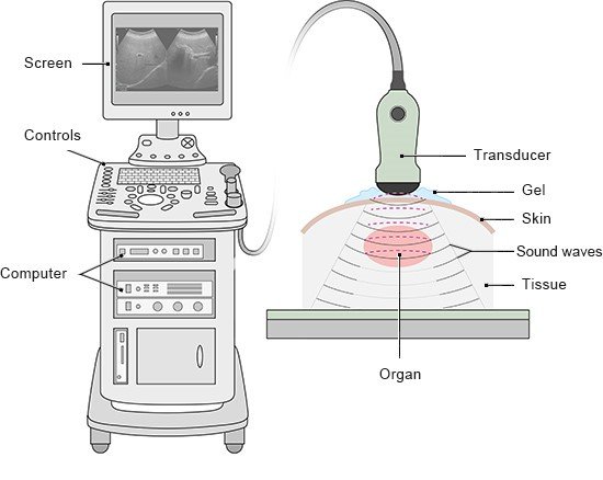
Niehaus J. Praxishandbuch Sonografie. Munich: Urban und Fischer; 2018.
Pschyrembel Online. 2024.
IQWiG health information is written with the aim of helping people understand the advantages and disadvantages of the main treatment options and health care services.
Because IQWiG is a German institute, some of the information provided here is specific to the German health care system. The suitability of any of the described options in an individual case can be determined by talking to a doctor. informedhealth.org can provide support for talks with doctors and other medical professionals, but cannot replace them. We do not offer individual consultations.
Our information is based on the results of good-quality studies. It is written by a team of health care professionals, scientists and editors, and reviewed by external experts. You can find a detailed description of how our health information is produced and updated in our methods.